천마의 망막신경세포 활성에 미치는 영향에 관한 연구

초록
천마를 사용한 신경 세포분화 실험과 항균실험을 실시하여 천마가 우리 눈에 미치는 영향을 알아보고자 하였다.
쥐의 망막신경세포주인 RGC5와 인간신경세포주인 SH-SY5Y를 대상으로 천마추출물의 농도별 세포생장률을 WST assay로 조사하고, ICC assay를 통해 분화에 미치는 영향을 confocal microscopy로 관찰하였다. 녹농균과 포도알균, 대장균에 대한 항균효과를 MIC assay와 well-diffusion 실험을 통해 조사하였다.
RGC5는 천마추출물 50-150 μg/ml에서, SH-SY5Y 세포주는 50-300 μg/ml 농도에서 세포독성이 없는 것을 확인하였다. 천마추출물 50, 100, 150 μg/ml에서 RGC5와 SH-SY5Y 세포주에 대하여 신경분화를 촉진시킴을 알 수 있었다. 또한 well-diffusion 결과 녹농균과 포도알균, 대장균에 대한 항균효과는 없는 것으로 나타났다.
천마추출물은 항균활성이 아닌 망막신경세포의 활성을 증가시킬 것으로 사료되는 바, 향후 천마추출물을 이용한 망막신경세포 활성 증가 방안의 연구를 통한 새로운 의, 약학적 적용연구가 진행되어져야 할 것이다.
Abstract
G. elata has been actively studied in the treatment of related diseases due to the efficacy of nerve protection and pain relief, but research on the effect of G. elata on our eyes never been explored. Therefore, we investigated on neuronal cell differentiation and antibacterial tests to fine the effect of G. elata on our eyes.
Cell cytotoxicity on retinal ganglion cell 5 (RGC5) and human neuroblastoma (SH-SY5Y) were determined by WST assay. ICC assay was used to observe the effect of the extracts on the differentiation by confocal microscopy. Additionally, the antibacterial effects of Pseudomonas aeruginosa, Staphylococcus aureus and Escherichia coli were measured by MIC assay and well-diffusion experiment.
As a result of the WST assay, it was confirmed that G. elata extracts was not cytotoxic at 50-150 μg/ml and 50-300 μg/ml on RGC5 and SH-SY5Y cell line, respectively. A result of the effects on the cell differentiation by ICC assay confirmed that RGC5 and SH-SY5Y cell lines were stimulated to differentiate to neuronal cells at 100, 150 μg/ml of G. elata extracts. It was found that there was no antimicrobial effect against P. aeruginosa, S. aureus and E. coli by well-diffusion.
G. elata extracts is thought to increase the activity of retinal neurons, not antibacterial activity. Therefore, new and pharmacological application studies should be carried out by studying the increase of retinal nerve cell activity using G. elata extracts.
Keywords:
Gastrodia elata, Eyes, Cell differentiation, Antibacterial effect키워드:
천마, 눈, 세포분화, 항균효과서 론
천마(天麻, Gastrodia elata Blume)는 한국, 일본, 중국 등에 자생하는 참나무에 버섯균(Armillaria ssp.)과 공생하여 자라는 다년생 난초과(Orchidaceae) 식물로써 뿌리가 없으며 괴경(덩이줄기)으로 자라는데,[1] 이를 쪄서 건조시킨 것이 여러 나라에서 많이 연구 되고 있는 생약 중 하나인 천마(Gastrodiae rhizoma)이다.[2] 천마의 뿌리는 가을에 수확하여 건조하여 사용하는데, 동양에서는 약 3000년 전부터 약재로 써왔고, 특히 치매, 두통, 스트레스 해소, 진통, 고혈압, 당뇨, 중풍, 기관지천식, 이뇨, 간질, 경련을 멈추게 하는 진경 등에 효과가 있어, 진통제, 구풍제, 쓸개즙 배출 촉진제, 진정제, 강장제 등 여러 용도로 사용되고 있다.[3] 천마는 한방에서 주로 많이 이용되는 약재로 이에 대한의, 약학적 응용과 관련된 국내외 연구가 급속한 진행을 보이고 있다. 그 중 신경보호 효능, 항혈관형성효과 및 항암효과, 진통효과, 항뎅기바이러스효과, 항혈전생성효과 등에 대한 연구가 가장 많이 되었고, 관련 질환으로는 알츠하이머, 간질, 경련 등이 있다.[4] 천마의 신경보호 효능에 대한 연구는 이미 보고되고 있는 바와 같이, 뇌실질출혈로 유발되는 신경세포 자연사를 억제하고,[5] 뇌부종과 신경세포의 세포독성 부종 억제효능이 있으며,[6] 치매 유발 독성에 대해 뇌신경세포 사멸을 보호하는 효과가 있음을 보여주었다.[7,8] 그러나 뇌신경세포와 밀접한 연관이 있는 우리 눈에 미치는 영향을 조사한 연구는 전무한 것으로 나타났다. 본 연구는 이러한 지속적인 연구과정의 일환으로 천마가 우리 눈에 미치는 영향을 알아보고자 쥐망막신경세포주인 RGC5와 신경세포 독성실험에 널리 활용되고 있는 Human neuroblastoma cell line인 SH-SY5Y의 분화에 미치는 영향을 조사하였다. 또한 시력교정용이나 미용적으로 콘택트렌즈를 착용하는 사람에게서 호발하고 있는 각결막염의 주원인균인 녹농균과 포도알균, 대장균에 대한 항균효과를 실험하였다.
대상 및 방법
1. 천마추출
Omniherb(Yeongcheon, Korea)에서 구입한 분말형태의 천마 50 g에 70% EtOH 500 ml를 가해 80oC에서 3시간 동안 중탕 가열한 후, Filter paper(Lot No.2)를 이용해 1차적으로 filter 처리하였다. 40oC에서 1시간동안 농축한 후 동결건조기를 이용하여 얻은 고형분을 cell culture water로 녹인 후 pH를 중성으로 보정하였다. 고형분의 무게와 비교해 최종 농도를 확인 한 후 사용하였다.[9]
2. 세포생장률
쥐의 망막신경세포주인 RGC5와 사람의 신경세포주인 SH-SY5Y는 1% 항생제와 10%의 fetal bovine serum (FSB; Gibco, USA)이 포함된 DMEM-low glucose (Gibco, USA) 배지와 DMEM-high glucose (Gibco, USA)를 각각 사용하여 37oC, 5% CO2 배양기에서 배양하였다.[10] 세포의 생장률은 Water Soluble Tetrazolium salt (WST; EZ-SOD assay kit, 두젠바이오) assay를 통해 확인하였다.[11] 각 cell을 96-well plate에 10×104 cell/ml로 분주하여 24시간 배양하고, 각기 다른 농도별 천마추출물을 처리한 후 24시간 배양하였다. 100 μl의 WST를 넣고 2시간 배양 후 ELISA reader(Tecan, Switzerland)를 이용하여 450 nm에서 흡광도를 확인하였다.
3. Immunocytochemistry(ICC)
6-well plate의 각 well에 cover glass를 넣고 60×104 CFU/ml의 농도로 cell seeding한 후 천마추출물을 농도별로 처리하여 충분히 반응하도록 한다. 3.5% formaldehyde로 cell 고정 후 PBS(Phosphate-buffered saline)로 충분히 세척한 다음, blocking buffer(1% BSA; Bovine serum albumin)에 30분간 담가둔 후, 1차 항체 (TUBB3), 2차 항체 (Alexa 488) 각각 한 시간씩 처리하고, PI(Propidium iodide)용액에 염색하였다. 매 처리과정마다 PBS로 세세히 닦아내고, 2차 항체처리 과정부터는 암실에서 진행하였다. 모든 처리가 끝나면 slide glass에 mounting medium을 떨어뜨리고 cover glass를 덮은 후 가장자리를 매니큐어로 봉입하여 제작된 표본을 confocal microscopy(Nikon, Japan)로 관찰하였다.[12]
4. 항균활성
최소저해농도(Minimal Inhibitory Concentration; MIC)를 결정하기 위해 Broth dilution method를 이용하였다.[13] 1×10⁶ CFU/ml의 Escherichia coli(ATCC 25922), Sthaphylococcus aureus(ATCC 25923), Pseudomonas aeruginosa(KCCM 11266)를 96-well plate에 100 μl씩 분주하고, 천마추출물을 각각 50, 100, 150 μg/ml의 농도별로 희석하여 100 μl씩 넣어 각각 4, 8, 24시간동안 배양하여 595 nm에서 흡광도를 측정한 후, 각 well의 균을 1×10³ CFU/ml가 되도록 희석하여 E. coli, S. aureus는 TSA (tryptic soy agar) 배지에, P. aeruginosa는 NA(Nutrient Agar) 배지에 50 μl씩 분주하여 24시간 후 colony counting을 시도하였다. 또한, 1×10⁵ CFU/ml의 각 균을 100 ml씩 배지에 도말한 후 직경 6 mm의 well에 10, 20, 50, 100, 200 μg/ml로 농도를 달리한 추출물을 50 μl씩 주입한 후 37캜의 incubator에서 24시간 배양하면서 well 주변의 clear zone (mm)을 측정하여 항균력을 조사하였다.
결과 및 고찰
1. 천마추출물이 RGC5와 SH-SY5Y세포주의 세포생존률에 미치는 영향
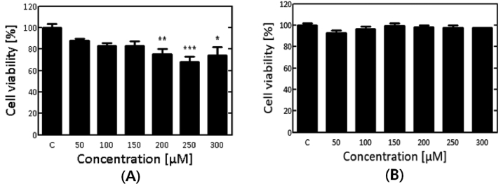
천마추출물이 신경세포에 독성이 있는지 확인하기 위하여 천마추출물에 대한 세포생존률을 평가하였다. 천마추출물의 세포생존률에 미치는 영향을 WST assay를 통해 확인한 결과는 Fig. 1과 같다. 쥐의 망막신경세포주인 RGC5의 경우 50-150 μg/ml에서 세포독성이 없음을 확인하였고, 200-300 μg/ml에서 약 20%의 세포생존률이 감소하는 것을 확인하였다(Fig. 1A). 사람의 신경세포주인 SH-SY5Y의 cell은 각 50-300 μg/ml 농도의 천마추출물에서 세포독성이 거의 없는 것을 확인 할 수 있었다(Fig. 1B). RGC5는 천마추출물 50-150 μg/ml에서 그리고 SH-SY5Y 세포주는 50-300 μg/ml 농도에서 세포독성이 없는 것을 확인하였다. 이 결과를 토대로 RGC5와 SH-SY5Y 세포주에서 세포독성이 없는 천마추출물 농도를 선별하여 본 실험에 적용하였다.
2. 천마추출물이 RGC5와 SH-SY5Y세포주의 세포분화에 미치는 영향
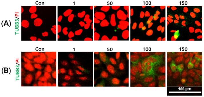
천마추출물이 RGC5와 SH-SY5Y세포주의 신경분화에 미치는 영향을 확인하였다(Fig. 2). RGC5세포의 경우 천마추출물 50-150 μg/ml에서 신경분화 마커인 TUBB3의 발현이 증가하는 것을 확인하였다(Fig. 2A). SH-SY5Y 세포도 마찬가지로 50-150 μg/ml에서 TUBB3의 발현이 증가하는 것을 확인하였다(Fig. 2B). 결과적으로 천마추출물 50, 100, 150 μg/ml에서 RGC5와 SH-SY5Y 세포주에 대하여 신경분화를 촉진시킴을 알 수 있었다. RGC5 세포의 경우 천마추출물의 신경분화에 대한 효능이 밝혀진 바 없으며, 본 연구 결과 천마추출물이 망막신경세포의 활성을 증가시킬 것으로 사료된다.

Effects of G. elata extracts on neuronal differentiation of RGC5 and SH-SY5Y cells. (A) indicate as RGC5, and (B) indicate as SH-SY5Y cell lines respectively. The concentration of G. elata extracts (1, 50, 100, 150 μg/ml) were treated for 24 hrs. Cells after culture were stained with PI staining solution. TUBB3 was visualized with fluorescence microscopy after immunofluorescence staining with TUBB3 antibody (green). Cell nuclei was shown in as for red color.
3. E. coli, S. aureus, P. aeruginosa에 대한 천마추출물의 항균활성
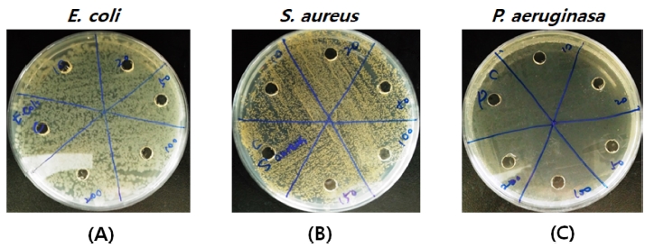
결막염 및 각막염의 대표적인 원인균인 세 가지 시험균주(E. coli, S. aureus, P. aeruginosa)를 선정하여 천마추출물에 대한 항균활성을 평가하였다. 천마추출물을 각각 50, 100, 150 μg/ml의 농도로 최소저해농도를 확인한 결과, 모든 시험균주(E. coli, S. aureus, P. aeruginosa)에 대하여 유의미한 항균활성도를 얻을 수 없었다(data not shown). 천마추출물의 농도 10, 20, 50, 100, 200 μg/ml에서 세 가지 시험균주(E. coli, S. aureus, P. aeruginosa)에 대한 well-diffusion 실험 결과, clear zone이 전혀 형성되지 않아 천마추출물의 항균활성은 없는 것으로 사료된다. 천마추출물의 항균활성 결과는 다음 Fig. 3과 같다. 이를 통하여 천마추출물은 항균활성이 아닌 신경세포 분화에 효능이 있음을 알 수 있었다.
4. 통계방법
모든 실험에 대한 측정치는 평균(mean)±표준오차(standard error of mean;SEM)로 Prism5의 One-way ANOVA Tukey test를 이용하여 통계처리 하였다. *p<0.5, **p<0.05, ***p<0.001은 통계적으로 유의함을 나타낸다.
결 론
본 연구는 천마가 우리 눈에 미치는 영향을 알아보고자 쥐망막신경세포인 RGC5와 신경세포 관련 실험에 널리 활용되고 있는 SH-SY5Y의 분화에 미치는 영향을 조사하였다. 그 결과, 천마추출물은 RGC5, SH-SY5Y의 두 세포 모두의 신경분화를 촉진시키는 것으로 나타났다. 또한 콘택트렌즈 착용자에서 호발하고 있는 각결막염의 주원인균인 녹농균과 포도알균, 대장균에 대한 항균효과 실험 결과, 천마추출물의 항균활성은 없는 것으로 나타났다. 따라서 천마추출물은 항균활성이 아닌 망막신경세포의 활성을 증가시킬 것으로 사료되는 바, 향후 천마추출물을 이용한 망막신경세포 활성 증가 방안의 연구를 통한 새로운 의, 약학적 적용연구가 진행되어져야 할 것이다.
References
-
Park, JP, Lee, SI, Jeong, JK., A Study on Functional components, antioxidant activity of Gastrodiae Rhizoma and Gastrodiae Elata floral axis, Kor J Herbology, (2015), 30(1), p19-24.
[https://doi.org/10.6116/kjh.2015.30.1.19.]

- The Ministry of Food and Drug Safety, Herbal Medicine Database, (2016), https://www.mfds.go.kr/herbmed/index.do?nMenuCode=7&code=KP-N35&includeUrl=/herbmed/view.jsp (25 December 2017).
- Huang, ZL., Recent developments in pharmacological study and clinical application of Gastrodia elata in China, Zhong Xi Yi Jie He Za Zhi, (1985), 5(4), p251-254.
-
Kim, HS, Lee, SI, Jeong, JK., Systemic review on the research trend of Gastrodiae Rhizoma and relationship between the herbology and KCD-code, Kor J Herbol, (2016), 31(2), p21-37.
[https://doi.org/10.6116/kjh.2016.31.2.21.]

- Song, SH, Lee, JS, Shin, JW, Kim, SJ, Sohn, NW., An immunohistochemical study on effect of Gastrodiae Rhizoma against neuronal apoptosis following intracerebral hemorrhage in rats, J Oriental Rehab Med, (2011), 21(4), p1-12.
- Lee, JY, Ku, JS, Lee, DE, Shin, JW, Kim, SJ, Sohn, NW., Effects of Gastrodiae Rhizoma on brain edema and aquaporin expressions following intracerebral hemorrhage in rats, Kor J Herbology, (2010), 25(4), p85-93.
-
An, H, Kim, IS, Koppula, S, Kim, BW, Park, PJ, Lim, BO, et al., Protective effects of Gastrodia elata Blume on MPP+-induced cytotoxicity in human dopaminergic SH-SY5Y cells, J Ethanopharmacol, (2010), 130(2), p290-298.
[https://doi.org/10.1016/j.jep.2010.05.006]

- Jung, YS, Kang, JH, Park, SH, Kwon, YM, Kim, GW, Koo, BS., Protective effect of Gastrodia elata on neuronal cell damage in Alzheimer's disease, J of Oriental Neuropsychiatry, (2010), 21(2), p125-140.
- Kim, HC, Lee, BN, Kim, YS, Lee, SI., Neuroprotective effect of Gastrodiae Rhizoma on dopaminergic neuronal cell death in Parkinson’s disease, Kor J Herbology, (1999), 14(1), p103-109.
-
Kim, IS, Koppula, S, Kim, BW, Song, MD, Jung, JY, Lee, G, et al., A novel synthetic compound PHID (8-Phenyl-6a, 7, 8, 9, 9a, 10-hexahydro-6H-isoindolo [5, 6-g] quinoxaline-7, 9-dione) protects SH-SY5Y cells against MPP(+)-induced cytotoxicity through inhibition of reactive oxygen species generation and JNK signaling, Eur J Pharmacol, (2011), 650(1), p48-57.
[https://doi.org/10.1016/j.ejphar.2010.09.063]

- Berridge, MV, Tan, AS, McCoy, KD, Wang, R., The biochemical and cellular basis of cell proliferation assays that use tetrazolium salts, Biochemica, (1996), 4(1), p14-19.
-
Bagasrawala, I, Zecevic, N, Radonjić, NV., N-Methyl D-Aspartate receptor antagonist Kynurenic acid affects human cortical development, Front Neurosci, (2016), 10, p435.
[https://doi.org/10.3389/fnins.2016.00435]

-
Balouiri, M, Sadiki, M, Ibnsouda, SK., Methods for in vitro evaluating antimicrobial activity: a review, J Pharm Anal, (2016), 6(2), p71-79.
[https://doi.org/10.1016/j.jpha.2015.11.005]


