
Effect of Endogenous Nitric Oxide Depletion on 12-Lipoxygenase Expression in Vascular Smooth Muscle Cells

Abstract
Nitric oxide (NO) plays a crucial role in maintaining retinal vascular homeostasis, and NO dysregulation is closely associated with retinal vascular diseases, such as diabetic retinopathy. We aimed to investigate whether the depletion of endogenous NO induces 12-lipoxygenase (12-LOX) expression in vascular smooth muscle cells (VSMCs) and to explore NO’s potential role in vascular dysfunction.
VSMCs were treated with the NO-scavenger PTIO as well as NO donors (SNAP and SNP) to modulate NO levels. Intracellular NO production was measured by fluorescence analysis. Using western blotting and RT-PCR, the expression of 12-LOX was evaluated at the protein and mRNA levels, respectively.
SNAP significantly increased NO production, whereas PTIO decreased NO levels in a concentration-dependent manner. PTIO-induced NO depletion significantly increased 12-LOX protein and mRNA levels. In contrast, co-treatment with SNAP or SNP attenuated the PTIO-induced upregulation of 12-LOX expression.
Endogenous NO negatively regulates 12-LOX expression in VSMCs. NO depletion induces 12-LOX expression, whereas the restoration of NO suppresses this effect. Therefore, the NO–12-LOX pathway plays an important role in retinal vascular dysfunction.
초록
산화질소(NO)는 망막 혈관 항상성을 유지하는 데 중요한 역할을 하며, 그 조절 이상은 당뇨망막병증과 같은 망막 혈관 질환과 밀접하게 관련되어 있다. 본 연구에서는 내인성 NO의 감소가 혈관평활근세포(VSMCs)에서 12-lipoxygenase(12-LOX) 발현을 유도하는지 규명하고, 이러한 변화가 혈관 기능 이상에 미치는 역할을 조사하고자 하였다.
VSMCs에 NO scavenger인 PTIO와 NO donor인 SNAP 및 SNP를 처리하여 NO level을 조절하였다. 세포 내 NO 생성은 형광 분석을 통해 측정하였으며, 12-LOX의 발현은 Western blot과 RT-PCR을 이용하여 각각 단백질 및 mRNA 수준에서 분석하였다.
SNAP 처리 시 NO 생성은 유의하게 증가한 반면, PTIO는 농도 의존적으로 NO 수준을 감소시켰다. PTIO에 의해 유도된 NO 감소는 12-LOX의 단백질 및 mRNA 발현을 유의하게 증가시켰다. 반면 SNAP와 PTIO를 함께 처리하였을 때 PTIO에 의해 증가된 12-LOX 발현은 억제되었다.
본 연구 결과는 내인성 NO가 VSMCs에서 12-LOX 발현을 음성적으로 조절함을 보여준다. NO의 감소는 12-LOX 발현을 유도하는 반면, NO의 회복은 이를 억제하여, NO-12-LOX 경로가 망막 혈관 기능 이상에 중요한 역할을 할 수 있음을 시사한다.
Keywords:
12-Lipoxygenase, NO, Diabetic retinopathy, VSMC키워드:
12-리폭시케나아제, 산화질소, 당뇨망막병증, 혈관평활근세포Introduction
Vascular dysfunction is a fundamental pathological feature of numerous cardiovascular and retinal diseases, including diabetic retinopathy, hypertension, and atherosclerosis. These conditions are characterized by abnormal vascular remodeling, increased vascular permeability, and dysregulated smooth muscle cell (VSMC) behavior, ultimately leading to impaired tissue perfusion and organ dysfunction.[1,2] Among the various molecular mechanisms underlying these pathological changes, redox imbalance and disruption of nitric oxide (NO) signaling have emerged as central contributors.[3]
NO is a critical regulator of vascular homeostasis. Produced primarily by endothelial nitric oxide synthase (eNOS), NO diffuses into VSMCs, where it promotes vasodilation through activation of the soluble guanylate cyclase (sGC)-cGMP pathway.[4] In addition to its vasodilatory function, NO exerts potent anti-inflammatory and anti-proliferative effects, inhibiting VSMC proliferation and migration while maintaining vascular quiescence.[5] Therefore, sufficient NO bioavailability is essential for preserving normal vascular function.
However, under pathological conditions such as oxidative stress, hyperglycemia, and inflammation, NO bioavailability is markedly reduced. This reduction occurs through multiple mechanisms, including decreased eNOS activity and increased consumption of NO by reactive oxygen species (ROS). In particular, superoxide rapidly reacts with NO to form peroxynitrite, a highly reactive oxidant that not only diminishes NO levels but also contributes to cellular damage and vascular dysfunction.[6,7] Thus, oxidative stress shifts the balance from a protective NO-dominant state to a pathological ROS-dominant state.
Lipoxygenases (LO) are a family of enzymes that metabolize arachidonic acid into bioactive lipid mediators, such as hydroxyeicosatetraenoic acids (HETEs), which play key roles in inflammation and vascular regulation.[8] Among these, 12-lipoxygenase (12-LOX) has been implicated in vascular pathologies through its metabolite, 12-hydroxyeicosatetraenoic acid (12-HETE). 12-HETE promotes VSMC proliferation and migration, enhances inflammatory responses, and contributes to endothelial dysfunction and increased vascular permeability.[9,10] These processes are closely associated with the progression of vascular diseases.
Emerging evidence indicates that NO and 12-LOX play opposing roles in vascular regulation, where NO exerts protective effects while 12-LOX promotes inflammatory and pathological vascular responses. Disruption of this balance is closely associated with retinal vascular diseases, such as diabetic retinopathy, which are characterized by NO deficiency, endothelial dysfunction, and abnormal vascular remodeling.[11,12] Nevertheless, the mechanistic link between endogenous NO depletion and 12-LOX expression has not been clearly established.
In this study, we tested the hypothesis that depletion of endogenous NO induces 12-LOX expression in VSMCs. To address this, we employed pharmacological modulation of NO levels using the NO scavenger PTIO and NO donors such as SNAP and SNP, and examined 12-LOX expression at both protein and mRNA levels. In addition, we examined whether restoration of NO levels using NO donors could reverse PTIO-induced 12-LOX expression. Through these approaches, we sought to clarify the mechanistic link between NO deficiency and 12-LOX-mediated vascular responses.
Materials and Methods
1. Cell culture and treatments
A10 cells (rat aortic smooth muscle cell line) were purchased from the American Type Culture Collection (ATCC, Manassas, VA, USA) and cultured in Dulbecco’s modified Eagle’s medium (DMEM; Sigma-Aldrich, St. Louis, MO, USA) supplemented with 10% fetal bovine serum (FBS), 100 U/ml penicillin, and 100 μg/ml streptomycin. Cells were maintained at 37°C in a humidified incubator with 5% CO2. For experimental procedures, cells were plated in 12-well plates at a density of 4×10⁴ cells per well and allowed to attach overnight. The medium was subsequently replaced with DMEM containing 0.5% FBS, and cells were pretreated with inhibitors for 30 min prior to exposure to PTIO under the indicated conditions.
2. Assay of NO formation
Intracellular nitric oxide (NO) production was measured using the fluorescent probe 4,5-diaminofluorescein (DAF-2; Sigma-Aldrich, St. Louis, MO, USA). DAF-2 reacts with NO to form a fluorescent triazolofluorescein, which emits green fluorescence upon excitation at 490–495 nm. A stock solution of DAF-2 (1 mg) was prepared in dimethyl sulfoxide (DMSO) and stored at −20°C. The working solution was prepared by diluting DAF-2 in 50 mM phosphate buffer (pH 7.4). VSMCs cultured in 12-well plates were treated with PTIO and/or NO donors (SNAP or SNP) as indicated. Cells were then incubated with DAF-2 (10 μM) for 30 min at 37°C. After incubation, cells were washed with phosphate-buffered saline (PBS), trypsinized, and collected on ice. Flow cytometric analysis was performed using a BD FACSLyric system (BD Biosciences, San Jose, CA, USA). Forward and side scatter parameters were used to gate viable cells, excluding debris. Fluorescence was detected in the FL-1 channel, and 10,000 events were acquired per sample. Data were analyzed as mean fluorescence intensity (MFI) and expressed as a percentage relative to the control group.
3. Protein analysis by Western blot analysis
Western Blot Analysis: VSMCs were lysed in RIPA buffer (Thermo Fisher Scientific, Waltham, MA, USA) containing protease inhibitor cocktail (Roche Diagnostics, Mannheim, Germany). Protein concentrations were determined using the Bradford assay (Bio-Rad, Hercules, CA, USA). Equal amounts of protein (30 µg) were separated by SDS-PAGE on 10% polyacrylamide gels and transferred onto polyvinylidene difluoride (PVDF) membranes (Millipore, Bedford, MA, USA). The membranes were blocked with 5% non-fat dry milk in Tris-buffered saline containing 0.1% Tween-20 (TBST) for 1 h at room temperature. The membranes were incubated overnight at 4°C with primary antibodies against 12-lip-oxygenase (12-LOX) (Abcam, Cambridge, UK) and β-actin (Cell Signaling Technology, Danvers, MA, USA) as a loading control. After washing with TBST, the membranes were incubated with horseradish peroxidase (HRP)-conjugated secondary antibodies (Cell Signaling Technology) for 1 h at room temperature.
4. Assay on Reverse Transcriptase-Polymerase Chain Reaction (RT-PCR)
(a) Reverse Transcription: First-strand cDNA was synthesized from 2 µg of total RNA using M-MLV reverse transcriptase (Promega, Madison, WI, USA). Total RNA was mixed with DEPC-treated water (Ambion, Austin, TX, USA) and 250 ng of random primers (Promega), heated at 75°C for 5 min, and chilled on ice for 5 min. Subsequently, 2 µl of 0.1 M DTT, 4 µl of 5× reaction buffer, 4 µl of 2.5 mM dNTP mixture (Thermo Fisher Scientific, Waltham, MA, USA), 100 U of reverse transcriptase, and 16.5 U of RNase inhibitor (Promega) were added. The reaction mixture was incubated at 37°C for 2 h, followed by heat inactivation at 100°C for 2 min. The synthesized cDNA was stored at −20°C until use.
(b) Polymerase Chain Reaction: PCR amplification was performed using specific primers for 12-lipoxygenase (12-LOX) and glyceraldehyde-3-phosphate dehydrogenase (GAPDH) as an internal control. PCR was carried out using Taq DNA polymerase (Takara Bio Inc., Shiga, Japan). The primer sequences were as follows: 12-LOX: Forward: 5′-TTCAAATGAGATTGTGGGAAAAT-3′, Reverse: 5′-AGATCATCTCTGCCTGAGTATCTT-3′, GAPDH: Forward: 5′-TGAAGGTCGGTGTGAACGGATTTGGC-3′, Reverse: 5′-CATGTAGGCCATGAGGTCCACCAC-3′. PCR products were separated on a 1.5% agarose gel (Invitrogen, Carlsbad, CA, USA) and visualized by ethidium bromide staining under UV illumination using a gel documentation system (Bio-Rad, Hercules, CA, USA).
5. Statistics
Statistical analysis was performed using one-way analysis of variance (ANOVA) followed by Tukey’s post hoc test for multiple comparisons using GraphPad Prism version 9.0 (GraphPad Software, San Diego, CA, USA). Data are presented as mean ± SEM, and differences were considered statistically significant at p < 0.05.
Results and Discussion
1. PTIO- and SNAP-mediated regulation of NO production in VSMCs.
PTIO was used as a nitric oxide (NO) scavenger to reduce endogenous NO bioavailability, whereas SNAP and SNP were employed as representative NO donors based on previous studies demonstrating their regulatory effects on NO signaling pathways.[13,14] To determine whether nitric oxide (NO) levels are modulated by pharmacological agents, vascular smooth muscle cells (VSMCs) were treated with the NO donor SNAP and the NO scavenger PTIO, and intracellular NO production was measured. As shown in Fig. 1, treatment with SNAP significantly increased NO levels compared with the control group (**p<0.01), confirming the effective generation of NO by SNAP. These findings are consistent with previous reports demonstrating that NO donors enhance intracellular NO bioavailability and regulate vascular function.[13,15]
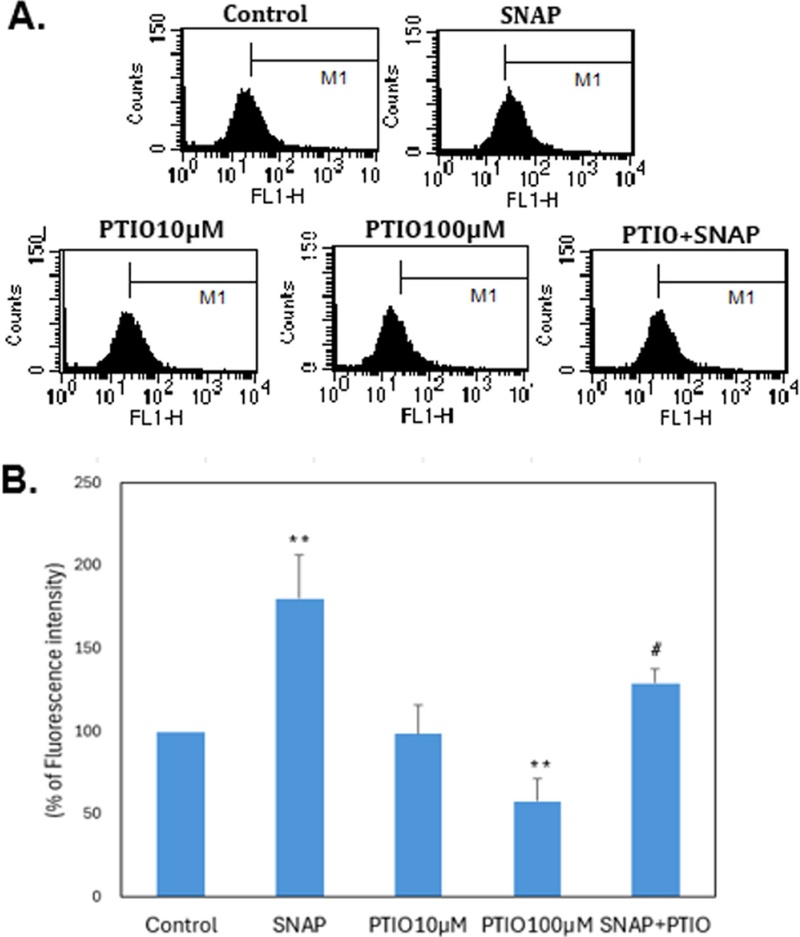
PTIO- and SNAP-mediated regulation of nitric oxide production in VSMCs. (A) Representative flow cytometry histograms showing intracellular NO levels in VSMCs treated with SNAP 50 µM (NO donor), PTIO (NO scavenger), or their combination. Fluorescence intensity (FL1-H) reflects intracellular NO levels. (B) Quantitative analysis of NO production expressed as a percentage of the control. VSMCs were treated with SNAP 50 µM (NO donor), PTIO (NO scavenger), or their combination. SNAP significantly increased NO production, whereas PTIO at 100 µM significantly decreased NO levels. Co-treatment with SNAP and PTIO attenuated SNAP-induced NO production. Data are presented as mean ± SEM (n=3). Statistical analysis was performed using one-way ANOVA followed by Tukey’s post hoc test. **p<0.01 vs. control; #p<0.05 vs. SNAP.
In contrast, treatment with PTIO reduced NO production in a concentration-dependent manner, with a significant decrease observed at 100 µM (**p<0.01 vs. control), indicating that PTIO effectively scavenges endogenous NO. PTIO has been widely used as a selective NO scavenger that reduces NO bioavailability in vascular cells.[13,16,17] Treatment with 10 µM PTIO showed no significant effect, suggesting that a threshold concentration is required to significantly deplete NO levels.
Importantly, co-treatment with SNAP and PTIO significantly attenuated SNAP-induced NO production (#p<0.05 vs. SNAP), demonstrating that PTIO effectively counteracts NO generated by SNAP. This result confirms that NO levels in VSMCs can be dynamically regulated by pharmacological manipulation. Given that NO is a critical regulator of vascular homeostasis and is closely associated with retinal vascular diseases, including diabetic retinopathy, modulation of NO levels may have significant pathological implications.[18,19] In particular, reduced NO bioavailability has been linked to endothelial dysfunction and abnormal vascular remodeling in retinal tissues.
Taken together, these results validate an experimental model of NO depletion and restoration in VSMCs, providing a basis for investigating the role of endogenous NO in regulating downstream targets such as 12-lipoxygenase (12-LOX).
2. PTIO-induced NO depletion increases 12-lipoxygenase protein expression in VSMCs.
To determine whether depletion of endogenous nitric oxide (NO) affects 12-LOX expression, VSMCs were treated with the NO scavenger PTIO, and 12-LOX protein levels were analyzed by Western blotting. As shown in Fig. 2A, PTIO treatment increased 12-LOX protein expression in a dose-dependent manner (10-200 µM), with maximal induction observed at 200 µM. Actin was used as a loading control.
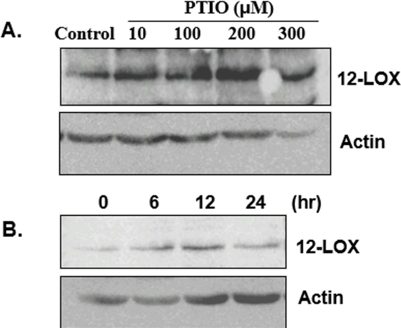
PTIO-induced NO depletion increases 12-lipoxygenase protein expression in VSMCs. (A) Representative Western blot analysis of 12-LOX protein expression in VSMCs treated with increasing concentrations of PTIO (10-200 μM). Actin was used as a loading control. (B) Time-dependent induction of 12-LOX protein expression in VSMCs following PTIO 100 μM treatment (0-12hr). Protein levels increased progressively over time. These results demonstrate that NO depletion induced by PTIO enhances 12-LOX protein expression in a dose- and time-dependent manner.
To further evaluate the temporal regulation of 12-LOX expression, VSMCs were treated with PTIO for different time periods. As shown in Fig. 2B, 12-LOX protein expression gradually increased over time, with a significant elevation observed at 12 and 24 h, indicating that NO depletion induces 12-LOX expression in a time-dependent manner. These findings demonstrate that reduction of endogenous NO is sufficient to induce 12-LOX protein expression in VSMCs. Given that NO is known to exert anti-inflammatory and anti-proliferative effects in vascular cells, its depletion may remove inhibitory constraints on pro-inflammatory signaling pathways, including lipoxygenase-mediated pathways.[20]
Previous studies have shown that 12-LOX plays a critical role in vascular inflammation and remodeling through the production of bioactive lipid mediators such as 12-HETE.[8] Upregulation of 12-LOX has been associated with increased smooth muscle cell proliferation, migration, and endothelial dysfunction, all of which contribute to the progression of vascular diseases. In particular, 12-LOX activation has been implicated in retinal vascular abnormalities, including diabetic retinopathy, where oxidative stress and NO deficiency are key pathogenic factors.[8,11]
Taken together with the findings from Fig. 1, these results suggest that NO depletion acts as an upstream regulatory signal that induces 12-LOX expression. This supports the concept that reduced NO bioavailability under pathological conditions may promote vascular dysfunction through activation of 12-LOX-dependent pathways.
3. PTIO-induced NO depletion increases 12-LOX mRNA expression in VSMCs.
To examine whether NO depletion regulates 12-LOX expression at the transcriptional level, VSMCs were treated with PTIO and 12-LOX mRNA was analyzed by RT-PCR. As shown in Fig. 3, PTIO increased 12-LOX mRNA expression in a concentration-dependent manner. These results indicate that NO depletion upregulates 12-LOX gene expression. Since NO suppresses inflammatory gene expression in vascular cells, its reduction may enhance transcription of 12-LOX. Increased 12-LOX expression is associated with vascular inflammation and remodeling and may contribute to retinal vascular diseases such as diabetic retinopathy.[21]
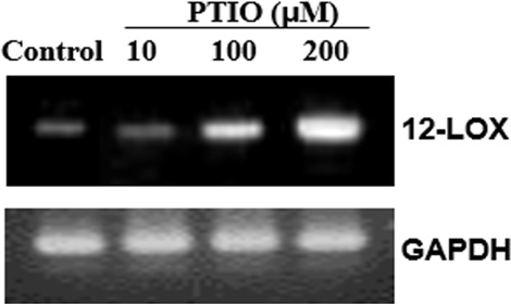
PTIO-induced NO depletion increases 12-LOX mRNA expression in VSMCs. Representative RT-PCR analysis of 12-LOX mRNA expression in VSMCs treated with various concentrations of PTIO (10-200 μM). GAPDH was used as an internal control. PTIO treatment increased 12-LOX mRNA expression in a dose-dependent manner, indicating transcriptional regulation of 12-LOX by NO depletion.
4. SNAP attenuates PTIO-induced upregulation of 12-LOX mRNA expression in VSMCs.
To determine whether restoration NO levels affects PTIO-induced 12-LOX expression, VSMCs were treated with PTIO in the presence or absence of NO donors (SNAP and SNP), and 12-LOX mRNA expression was analyzed by RT-PCR. As shown in Fig. 4, PTIO treatment markedly increased 12-LOX mRNA expression, whereas co-treatment with SNAP or SNP significantly attenuated this effect.
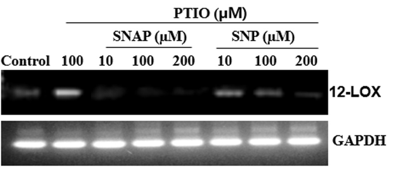
SNAP attenuates PTIO-induced upregulation of 12-LOX mRNA expression in VSMCs. Representative RT-PCR analysis showing 12-LOX mRNA expression in VSMCs treated with PTIO in the presence or absence of NO donors (SNAP or SNP). GAPDH was used as an internal control. PTIO-induced upregulation of 12-LOX mRNA expression was markedly reduced by co-treatment with SNAP or SNP, indicating that restoration of NO levels suppresses 12-LOX expression. PTIO-induced upregulation of 12-LOX mRNA expression was markedly reduced by co-treatment with SNAP or SNP, indicating that restoration of NO levels suppresses 12-LOX expression.
These results indicate that restoration of NO levels suppresses PTIO-induced upregulation of 12-LOX, suggesting that NO negatively regulates 12-LOX expression. NO is known to inhibit inflammatory signaling and gene expression in VSMCs,[22] and its depletion may therefore promote activation of pro-inflammatory pathways, including lipoxygenase signaling.
Previous studies have demonstrated that 12-LOX is upregulated in vascular smooth muscle cells under inflammatory and proliferative conditions and contributes to vascular remodeling and dysfunction.[23] In addition, 12-LOX-derived metabolites have been implicated in the regulation of vascular tone and NO signaling, indicating a reciprocal relationship between NO and lipoxygenase pathways.[24] Although the precise mechanism by which NO depletion induces 12-LOX expression remains unclear, accumulating evidence suggests close interactions between NO signaling and the lipoxygenase pathway. Ma et al. reported that NO regulates arachidonic acid metabolism and modulates the generation of 12-LOX-derived metabolites, supporting a functional link between NO signaling and lipoxygenase activity.[25] Furthermore, reduced NO bioavailability has been associated with enhanced 12-LOX expression in vascular smooth muscle cells under pathological conditions.[26]
Based on these findings and the present results, it is possible that depletion of endogenous NO removes inhibitory regulation on 12-LOX expression, thereby promoting 12-LOX-mediated vascular responses. Conversely, restoration of NO levels suppresses 12-LOX expression, supporting the concept that NO acts as a negative regulator of 12-LOX-mediated vascular dysfunction.
Conclusions
In conclusion, depletion of endogenous nitric oxide (NO) induces 12-lipoxygenase (12-LOX) expression in VSMCs, whereas restoration of NO suppresses this effect. These findings suggest that NO functions as a negative regulator of 12-LOX expression and that reduced NO bioavailability may promote vascular dysfunction through lipoxygenase-associated inflammatory signaling. Given the involvement of NO deficiency and lipoxygenase pathways in retinal vascular diseases such as diabetic retinopathy, the NO-12-LOX axis may represent a novel mechanism underlying retinal vascular dysfunction and a potential therapeutic target for oxidative stress-associated retinal vascular diseases.
Acknowledgments
This work was supported by the Suseong University Research Grant in 2025.
References
-
Beckman JA, Creager MA. Vascular complications of diabetes. Circ Res. 2016;118(11):1771-1785.
[https://doi.org/10.1161/CIRCRESAHA.115.306884]

-
Cheung N, Mitchell P, Wong TY. Diabetic retinopathy. Lancet. 2010;376(9735):124-136.
[https://doi.org/10.1016/S0140-6736(09)62124-3]
-
Förstermann U, Sessa WC. Nitric oxide synthases: regulation and function. Eur Heart J. 2012;33(7):829-837.
[https://doi.org/10.1093/eurheartj/ehr304]
-
Man AWC, Zhou Y, Xia N, et al. Endothelial nitric oxide synthase in the perivascular adipose tissue. Biomedicines. 2022;10(7):1754.
[https://doi.org/10.3390/biomedicines10071754]
-
Lamb FS, Choi H, Miller MR, et al. Vascular inflammation and smooth muscle contractility: the role of Nox1-derived superoxide and LRRC8 anion channels. Hypertension. 2024;81(4):752-763.
[https://doi.org/10.1161/HYPERTENSIONAHA.123.19434]
-
Lee JY. The role of 4-hydroxynonenal for activation of vascular smooth muscle cell. Korean J Vis Sci. 2019;21(2): 281-289.
[https://doi.org/10.17337/JMBI.2019.21.2.281]
-
Förstermann U. Oxidative stress in vascular disease: causes, defense mechanisms and potential therapies. Nat Clin Pract Cardiovasc Med. 2008;5(6):338-349.
[https://doi.org/10.1038/ncpcardio1211]
-
Funk CD. Prostaglandins and leukotrienes: advances in eicosanoid biology. Science. 2001;294(5548):1871-1875.
[https://doi.org/10.1126/science.294.5548.1871]
-
Natarajan R, Nadler JL. Lipoxygenases and lipid signaling in vascular cells in diabetes. Front Biosci. 2003;8(6):783-795.
[https://doi.org/10.2741/1144]
-
Chen S, Zou H. Key role of 12-lipoxygenase and its metabolite 12-hydroxyeicosatetraenoic acid (12-HETE) in diabetic retinopathy. Curr Eye Res. 2022;47(3):329-335.
[https://doi.org/10.1080/02713683.2021.1995003]
-
Kowluru RA, Chan PS. Oxidative stress and diabetic retinopathy. Exp Diabetes Res. 2007;43603:1-12.
[https://doi.org/10.1155/2007/43603]
-
Moncada S, Higgs A. The L-arginine-nitric oxide pathway. N Engl J Med. 1993;329(27):2002-2012.
[https://doi.org/10.1056/NEJM199312303292706]
-
Millar CGM, Thiemermann C. Carboxy-Ptio, a scavenger of nitric oxide, selectively inhibits the increase in medullary perfusion and improves renal function in endotoxemia. Shock. 2002;18(1):64-68.
[https://doi.org/10.1097/00024382-200207000-00012]
-
Moncada S, Higgs A. The L-arginine-nitric oxide pathway. N Engl J Med. 1993;329(27):2002-2012.
[https://doi.org/10.1056/NEJM199312303292706]
-
Kingma JG Jr, Simard D, Rouleau JR. Nitric oxide bioavailability affects cardiovascular regulation dependent on cardiac nerve status. Auton Neurosci. 2015;187:70-75.
[https://doi.org/10.1016/j.autneu.2014.11.003]
-
Lee J, Kang S, Lee S. Expression of cyclooxygenase-2 by 4-hydroxynonenal in vascular smooth muscle cells. J Korean Ophthalmic Opt Soc. 2025;30(4):413-419.
[https://doi.org/10.14479/jkoos.2025.30.4.413]
-
Lee J. Induction of 5-lipoxygenase by 4-hydroxynonenal via nitric oxide generation in vascular smooth muscle cells. J Korean Ophthalmic Opt Soc. 2023;28(3):255-262.
[https://doi.org/10.14479/jkoos.2023.28.3.255]
-
Opatrilova R, Kubatka P, Caprnda M, et al. Nitric oxide in the pathophysiology of retinopathy: evidences from preclinical and clinical researches. Acta Ophthalmol. 2018;96(3):222-231.
[https://doi.org/10.1111/aos.13384]
-
Chen S, Zou H. Key role of 12-lipoxygenase and its metabolite 12-hydroxyeicosatetraenoic acid (12-HETE) in diabetic retinopathy. Curr Eye Res. 2022;47(3):329-335.
[https://doi.org/10.1080/02713683.2021.1995003]
-
Shin WS, Hong YH, Peng HB, et al. Nitric oxide attenuates vascular smooth muscle cell activation by interferon-gamma: the role of constitutive NF-κB activity. J Biol Chem. 1996;271(19):11317-11324.
[https://doi.org/10.1074/jbc.271.19.11317]
-
Natarajan R, Rosdahl J, Gonzales N, et al. Regulation of 12-lipoxygenase by cytokines in vascular smooth muscle cells. Hypertension. 1997;30(4):873-879.
[https://doi.org/10.1161/01.hyp.30.4.873]
-
Grootaert MOJ, Moulis M, Roth L, et al. Vascular smooth muscle cell death, autophagy and senescence in atherosclerosis. Cardiovasc Res. 2018;114(4):622-634.
[https://doi.org/10.1093/cvr/cvy007]
-
Kulkarni A, Nadler JL, Mirmira RG, et al. Regulation of Tissue Inflammation by 12-Lipoxygenases. Biomolecules. 2021;11(5):717.
[https://doi.org/10.3390/biom11050717]
-
Gliozzi M, Scicchitano M, Bosco F, et al. Modulation of nitric oxide synthases by oxidized LDLs: role in vascular inflammation and atherosclerosis development. Int J Mol Sci. 2019;20(13):3294.
[https://doi.org/10.3390/ijms20133294]
-
Ma Z, Ramanadham S, Corbett JA, et al. Interleukin-1 enhances pancreatic islet arachidonic acid 12-lipoxygenase product generation by increasing substrate availability through a nitric oxide-dependent mechanism. J Biol Chem. 1996;271(2):1029-1042.
[https://doi.org/10.1074/jbc.271.2.1029]
-
Kim HY, Jeong DW, Park HS, et al. Comparison of 12-lipoxygenase expression in vascular smooth muscle cells from old normotensive Wistar-Kyoto rats with spontaneously hypertensive rats. Hypertens Res. 2013;36(1):65-73.
[https://doi.org/10.1038/hr.2012.119]