독일식과 영미식 주시시차에 대한 방법론적 분석

초록
명실과 반암실에서의 안위변화를 알아보고 독일식 양안시 검사법 MKH와 영미식 검사법에 따라 주시시차를 측정하고 비교한다.
안질환이 없으며 교정시력 0.8 이상인 다양한 연령대의 성인 35명을 대상으로 MKH를 실시했다. MKH는 전형적인 절차를 따르지 않고 본 연구 목적에 적합하게 수정되어 사용되었다.
조도의 변화에 따라 전체 35명 중 6명(30세 이하)에게서 변화가 나타났으며 그 중 4명은 어두울수록 내전 경향을 보였고 2명은 외전 경향을 보였으나 샘플 수가 작아 통계적 의미를 확인할 수 없었다. 독일식 검사법에 의한 주시시차는 15.7±12.4', 영미식 방법에 의하면 65.4±52.8'로 나타나 분명한 차이가 있는 것으로 나타났다(p=0.001).
검사실에서의 조명은 젊은 연령대에서의 안위에 변화를 주는 경향이 있다고 생각되며 독일식 및 영미식 주시시차는 논리적 측정방법에서 상호 큰 차이가 있는 것으로 나타났다.
Abstract
The present study aimed to investigate ocular deviation in bright and ambient rooms and compare the fixation disparity using the German binocular measurement methodology Mess und Korrektionsmethodik nach Haase(MKH) to the one based on the idea of anglosphere.
Thirty-five subjects from the adult age group involving no specific ocular disease and greater than 0.8 visual acuity participated in the study. The conventional MKH in Germany was modified for this study.
There was a change in ocular deviation in 6 subjects (age ≤30 years), depending on the change in the luminance in the room. Four of those showed adduction and two showed abduction in tendency, which was not statistically analyzed because of the lack of samples. There was a significant difference between the fixation disparity by MKH and the one by the idea of anglosphere (15.7±12.4' and 65.4±52.8', respectively; p=0.001).
It is considered that the change in luminance in a room has a tendency of unstable ocular deviation especially in younger people. There is a quantitatively huge difference between the anglosphere and German logical methodologies in measure.
Keywords:
MKH, Ocular deviation, Fixation disparity, Motor fusion, Sensory fusion키워드:
MKH, 안위, 주시시차, 운동성융합, 감각성융합서 론
양안 망막결상의 좌표 값이 서로 다른 값을 갖는 경우는 첫째, 사물의 거리감을 발생시키는 비대응결상이거나 둘째, 좌표 값 간 차이가 작아 융합버전스가 작동되어 양안단일시를 이루기도 하지만 차이가 임계점을 넘어서서 복시를 발생시키는 이상대응결상을 하는 경우를 들 수 있다. 전자의 경우 양질의 입체시생활을 하는 데에 반드시 필요한 시각체계이지만 후자의 경우에 해당하는 이상망막 대응이 전자에 해당하는 입체시생활을 방해할 수도 있다.
양안시 영역에서 중요하게 다루어지는 요소 중 하나인 주시시차(fixation disparity, 이하 FD)는 양안의 시축간 버전스 각이 다르지만 양안단일시가 유지되는 상태를 말한다. 이것은 이상망막대응(anomalous retinal correspondence)하는 양안의 두 좌표 값이 서로 적응하여 감각적으로 융합할 수 있는 가상의 영역 내에 존재하기 때문이며 이 가상의 영역을 파눔 영역(Panum’s fusional area)이라고 한다. 파눔 영역에 속해 있는 두 개의 이상망막대응 좌표가 이루는 각은 일반적으로 6'(minutes of arc)이하로 작지만 크게는 30'(약 1 prism diopter, Δ)까지도 나타날 수 있다.[1] 원칙적으로, FD는 상호 적응에 의해 단일시를 이루지만 입체시각 및 버전스 운동 기능을 저하시키며[2-3] 나아가 안정피로를 야기하는 등 시생활의 질을 떨어뜨릴 수 있는 요인으로 임상적인 처치를 필요로 한다.[4]
흥미롭게도 FD는 나라별로 각각 다른 관점과 해석을 보이는데 최근 연구 자료에 의하면 영미식 FD는 프리즘 부가에 따른 운동성 버전스 요구량에 따라 곡선의 y축과의 절편으로 정의하는 한편 독일식 FD는 양안의 편위량이 감각적으로 적응하지만 그 양이 증가하게 될 때 비로소 발현되는 것으로 해석한다.[5] 가령, 영미식 FD는 6 m 거리의 양안융합역이 존재하는 시표를 대상으로 3 cm의 편차를 의식한다면 약 17'으로 표현하며, 독일식 FD는 먼저 완전융합제거사위검사의 일환인 십자시표 검사에서 나타난 2 Δ를 교정한 이후 일부융합제거사위검사인 시계침시표에서 추가로 나타난 0.5 Δ만을 분각으로 환산한 약 17'만을 FD로 간주한다. 요컨대, 독일어권에서는 중앙에 양안고시점이 없는 십자시표를 먼저 사용하여 외안근의 융합력을 측정 및 교정한 이후 양안고시점이 있는 시계침시표 등을 사용하여 잔존하는 이상량을 FD로 나타내는 반면, 영어권에서는 외안근의 융합력을 아직 교정하지 않은 채 처음부터 곧장 중앙에 양안고시점이 있는 시표를 사용하여 FD 측정을 하는 것이 대표적인 큰 차이점이라고 할 수 있다.
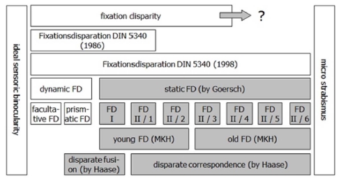
융합을 위한 외안근의 부담을 프리즘으로 덜어준 이후 추가로 발생하는 안위를 FD로 규정하고 그에 따라 검사 방법을 고안한 독일식 양안시 검사법 이른바 MKH(Messund Korrektionsmethodik nach Haase; 하제식 교정 방법론)에 따르면 FD를 교정효과를 볼 수 있는지 여부에 따라 세부적으로 분류했다(Fig. 1). 그의 이론에 따르면 FD가 형성되지 오래되지 않은 경우에는(FD I와 FD II/1-2) 프리즘 교정시 그에 따른 교정효과를 볼 수 있는데 반해 FD가 형성된 지 상당한 기간이 지난 경우에는(FD II/3-6) 이미 한쪽 눈의 중심와에서 억제현상이 발생해 프리즘 교정으로 큰 효과를 볼 수 없거나 오히려 역효과가 나타날 수 있다고 강조한다(Fig. 2).
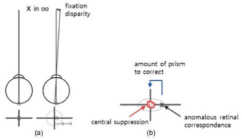
Suppressed fovea by FD in Panum’s area (Reprinted with kind permission from Yoon JH and Seo JM).[7] (a) The binocular single vision still remains despite the abduction of right eye because the retinal image of the right eye is so in Panum’s area that the visual excitations from different coordinates are unified. FD presences when the fovea of one eye is slightly misaligned from pointing at the binocularly viewed target, (b) Once fixation disparity occurs the foveal area becomes inactivated and gradually degraded in terms of visual function. It ultimately results in suppression of the eye with FD.
MKH를 사용하여 이번 연구를 처음 시작한 본 연구진은 독일식 FD의 실체적 확인뿐만 아니라 Haase에 따른 FD 등급의 분류 나아가, MKH의 궁극적 목적인 입체시의 질적 개선 그리고 그 이후에 대한 연구를 연속적으로 진행하고자 한다. 본 연구에서는 먼저 명실에서 반암실로 검사실 환경이 바뀔 때 안위 결과에서 어떤 변화가 나타나는지 확인하고자 한다. 한국의 임상 현장에서 사용하는 양안시 검사장비는 대부분 빔프로젝터 방식을 사용하기 때문에 검사실 환경이 반암실 형태를 벗어나기 어렵다. 게다가 우리는 시작업에 할애하는 대부분의 시간을 주간에 사용하고 있기 때문에 괴리는 분명히 존재하며 이미 많은 연구에서 밝혀진 바 있다.[8-13] 그러나 안위의 방향이나 그 정도가 모두 다르기 때문에 명실에서도 사용이 가능한 발광형 검사 장비인 Polatest를 사용하여 검사실의 밝기가 안위에 어떤 영향을 주는지 알아보고자 한다. 또한 영미권에서 사용하는 FD 측정이론에 의한 방법과 독일어 권역(독일, 오스트리아, 네델란드, 스위스)에서 사용하는 FD 측정 방법에 따라 결과 값을 비교해보고 그 의미를 해석하여 임상에서 경험하는 FD 측정 기준이나 방법에 대한 혼란을 줄이는 데에 조그마한 보탬이 되고자 한다. 나아가 주시시차와 관련한 국내의 연구들은 영미식 검사장비와 독일식 검사장비를 각각 활용하여 결과를 비교했으나[14,15] 그들 간 검사방법론적 고찰이나 장비의 통일성을 이루지 못한 부분에 대한 도전적 시도로 본 연구의 작은 의의를 두고자 한다.
대상 및 방법
본 연구의 취지에 동의한 사람 가운데 전신질환이나 안질환이 없으며 교정시력이 0.8 이상인 성인(40.46±18.97세) 남녀 35명(남자 22명, 여자 13명)을 대상으로 했다. 약도(1.5 Δ이하)의 수직방향의 편위를 가진 사람은 두 명이었으나 본 연구에서는 수평 FD에 중점을 두었기 때문에 연구 결과에서 제외했다. 완전 교정된 대상자는 초기 FD 검사에 주로 사용되는 십자시표와 시계침 시표 검사를 하여 그 결과 값을 분석에 사용했다. 밝은 장소와 어두운 장소에서의 안위 변화를 알아보기 위해 발광형 검사시표인 i.Polatest® (version 1.2 by Carl Zeiss Vision GmbH, Aalen, Germany)를 사용하였다. 모니터의 크기는 299.5 mm × 223.5 mm 이며 주사율은 60Hz였다. 검사실의 조명을 끄고(50~60 lux) 켠 채(1300~1500 lux) 안위의 변화를 측정했다. 암소시 상태에서의 안위 변화를 알아보기 위해 약 10 분의 사전 적응시간이 주어졌다. 모든 대상자는 Polatest와 일반적으로 같이 사용되는 시험테(Oculus trial frame, Oculus, Germany)를 사용하여 검사거리 5 m에서 전체 검사를 진행했다.
Haase가 개발한 MKH의 검사 진행방식에 따르면 십자시표 검사의 본래 목적은 자연시 상태에서 양안융합역을 최소화 시킨 시표로서 외안근에 의한 융합 정도를 측정하기 위함이며 시계침 시표 검사와 ‘ㄷ’자 시표는 양안융합역을 포함시켜 신경상의 적응도를 측정하기 위함에 있다. 다만 본 연구에서는 명소시와 암소시에서의 안위변화 그리고 주시시차 측정을 위해 운동성 융합에 필요한 프리즘을 교정하기 전과 후 처방 값에 있어서 어떤 변화가 있는지 살펴보고자 연구를 다음과 같이 설계했다(Fig. 3). 먼저 영미식 주시시차 측정을 위해 외안근의 융합력을 제시해주는 십자시표 검사를 하지 않고 곧장 시계침시표에서 측정 및 교정량을 기록했다(Fig. 3(c)). 독일식 주시시차 측정은 십자시표에서 측정 및 교정을 한 이후 시계침시표에서 추가로 측정된 양만을 주시시차로 간주하여 이 두가지 방법에 의한 결과를 비교 분석에 활용했다(Fig. 3(b)).
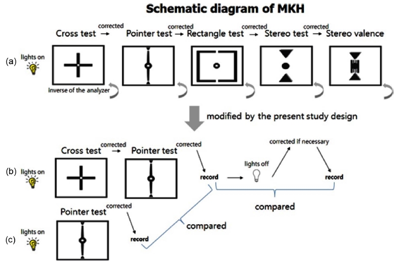
Modified MKH for the present study (Reprinted and modified with kind permission from Yoon JH and Seo JM)[7]; (a) MKH should be conducted in order above to correct the ‘Winkelfehlsichtigkeit’ (associated phoria translated in English). Correction with the prism in every single test ends and the axes of polarization should be converted to check whether there is any change in ocular deviation, (b) German fixation disparity. In order to meet the study purpose the tests after the Pointer test are not conducted. Instead, it is investigated to see if there were any changes between lights on and off, (c) Anglo-American fixation disparity. Without correction in the Cross test it directly goes to the Pointer test after the refraction. After correction in the Pointer test the results are compared to the ones obtained from (b) under the lights on.
결과 및 고찰
본 연구에 참가한 대상자는 최고령인 77세부터 20세까지로 총 35명을 대상으로 했다. 20대에서와 60대 이후의 고령에서 외사위의 밀집현상을 보였으며 안위이상은 나이에 따라 음의 상관관계가 있는 것으로 나타났다(r = −0.3).
검사실 밝기에 따른 안위의 변화를 알아보기 위해 일반적인 명소시 조건 하 시계침 시표에서 프리즘 교정 직후 검사실의 조명을 차단시켜 반암실 조건으로 전환시켜 안위의 변화량을 살피면서 정위에 필요한 양만큼 재 교정했다. 전체 35명 중 6명에게서 변화가 나타났으며 이들 모두 30세 이하인 것으로 나타났다(Fig. 4). 조도의 변화에 따라 안위가 변한 6명 중 4명은 내전 경향(0.63±0.22 Δ)을 보였으며 2명은 외전 경향(0.75±0.25 Δ)을 보였으나 작은 샘플 수로 인해 통계적 의미는 확인할 수 없었다.
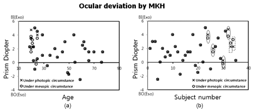
(a) Ocular deviation with increasing age under photopic and mesopic circumstance. For the younger subjects it appeared to be stronger in sensitivity to the change of lights, (b) It was hard to identify the changes between the lights on and off with plotting (a) and replotted. Those who had a change in ocular deviation were 6 in total. Two dashed rectangles represent the subjects with the adduction and four solid ellipses represent the subjects with the abduction in tendency.
보통 자율신경계에 의해 지배되는 섬모근은 핀홀, 암소시, 지속적인 동일 자극 등에 의해 시각피드백 회로(visual feedback loop)를 작동시킨다고 알려져 있다.[8] 특히, 암소시에 개인별로 큰 편차를 가지나(0~4 D) 평균 1.5 D까지 근시화 된다고 알려져 있으며[9] 그 현상은 나이에 따라 그 정도가 감소하는 경향이 있으며[10] 정시나 원시에 비해 근시에서 정도가 낮게[11] 나타나기 때문에 야간근시는 결국 굴절이상도와 밀접하게 연관되었음을 알 수 있다.[12] 이렇게 형성된 야간근시는 외전 경향을 보이는 것으로 알려져 있으나 Hilz는 광투과율을 조정할 수 있는 필터렌즈를 사용하여 마독스간과 폴라테스트로 사위를 측정한 결과 어두울수록 마독스간이나 폴라테스트 모두 내전하는 경향을 보였으나 그 양이나 방향성은 개인차가 커서 일반화시키기 어렵다고 보고했다.[13]
외안근에 의한 안위 보정량 즉, 운동성 융합 부분에서는 전체 대상자 중 30명(85.7%)이 외사위를 보였으며 평균 1.73±1.24 Δ으로 나타났다(Fig. 5). 양안융합역이 없는 십자시표에서 운동성 융합량을 교정 후 곧장 시계침 시표를 사용하여 신경 적응에 의한 안위 보정량 즉, 감각성 융합부분을 측정했는데 전체 35명 중 65.7%인 23명에서 0.36±0.38 Δ(12.2'±12.9')으로 나타났다. 한편, 운동성 융합부분에서 내편위를 보인 사람은 총 5명으로 나타났으며 그 중 한 사람만 시계침시표에서 0.5 Δ 외편위가 발생하여 내사위를 가지면서 외방 FD가 있는 사람은 한 사람으로 나타났다.

Prevalence and type of ocular deviation for all subjects. The motor portion (A) was measured in the Cross test without central fusion lock, which stands for the total magnitude of movement by extraocular muscles to keep binocular single vision. The sensor portion after correcting (A) implies the measurement of ocular deviation in the Pointer test with central fusion lock after correcting in the Cross test.
먼저 운동성융합 부분을 교정한 후 FD를 측정하는 독일식 검사법에 의하면 15.7±12.4'이 측정되었으며 운동성융합 부분을 교정하지 않은 채 곧장 FD를 측정하는 영미식 방법에 의하면 65.4±52.8'이 측정되어 FD를 해석하는 관점에 따라 분명한 차이가 있는 것으로 나타났다(Fig. 6(a), p=0.001). 약 34'은 1 Δ로 환산할 수 있기 때문에 영미식과 독일식 검사법에 의해 측정된 FD의 차이인 약 50 '은 1.5 Δ 정도라고 할 수 있으며 이 정도의 값은 FD의 일반적인 범위에 비춰볼 때 대단히 큰 값에 속한다. 이미 서론에서 두 가지 FD의 방법론적 차이를 설명한 바와 같이 영미식에서는 운동성 융합부분을 교정하지 않은 채 곧장 주시시차를 측정했기 때문에 운동성 융합역까지 포함된 값인 65.4±52.8'이 측정되었고 독일식에서는 운동성 융합부분을 선 교정 후 감각석 융합역의 편차만 측정했기 때문에 15.7±12.4'으로 낮게 측정되었다고 볼 수 있다. 이것은 아직 교정되지 않고 존재하던 외안근의 긴장이 파눔 영역권까지 영향력을 행사하여 발생한 감각적 적응도 즉, FD의 크기에 영향을 주었기 때문으로 풀이된다. 또한 운동성 융합 부분과 감각성융합 부분을 합한 정렬프리즘 값과 감각성융합을 단독으로 측정한 정렬프리즘 값을 비교해 본 결과 역시 각각 2.4±1.8 Δ와 1.9±1.6 Δ으로 나타나 검사의 방법론적 비교에서도 차이가 있는 것으로 나타났다(Fig. 6(b), p=0.015).
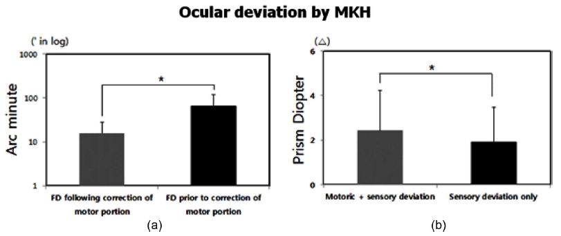
(a) Contrast between FD following correction of motor portion based on MKH and FD prior to correction of motor portion based on the methodology in anglosphere. There was a significant difference (p=0.001). (b) The deviation by MKH (motor + sensory deviation) was not significantly same the one by the methodology in anglosphere (sensory deviation only, p=0.015).
한편, 프리즘 분리법과 MKH를 사용하여 비교 분석한 최근의 국내 연구결과에 의하면 두 가지의 검사에 의한 프리즘 처방 간 큰 차이가 없었다고 보고했으나(p=0.345) 연구 방법에 있어서 오류가 있다고 생각된다.[14] 완전융합제거사위만을 교정하여 자각적 증상완화를 목표로 하는 미국의 프리즘 분리법(von Graefe technique)은 완전융합제거사위뿐만 아니라 일부융합제거사위를 모두 교정하며 특히, FD의 교정여부와 입체시의 질적 개선을 주목적으로 개발된 독일의 MKH와 근본적인 목적을 달리하고 있어서 상호 비교대상이 될 수 없기 때문이다. 그럼에도 불구하고 본 연구에서 성격이 다른 영미식 FD 검사와 독일식 FD 검사 간 비교를 할 수 있었던 이유는 동일한 검사 장비로 두 검사의 대표적인 이론적 논리만을 취하여 비교했기 때문에 결과 분석에 있어 타당성이 성립되었다고 생각된다.
결 론
FD를 이해하게 된지 아직 한 세기도 채 되지 않은 만큼 해석하고 바라보는 관점들이 다양하게 존재해 왔다. 본 연구에서는 검사실 조명이 시표의 선명도에 영향을 주지 않는 발광형 시표를 사용하여 검사실 조명의 점등 여부와 영미식 FD와 독일식 FD의 차이를 알아보고자 하였다. 검사실에서의 조명은 안위에 변화를 주는 것으로 나타났다. 20대에서 70대 후반의 연령대를 대상으로 한 본 연구에서는 20대 연령에서만 안위의 변화가 있는 것으로 나타났다. 그러나 안위의 방향성이나 그 정도는 해당되는 대상자가 많지 않아 의미 있는 결과를 제시할 수 없었던 점이 본 연구의 한계로 남게 되었다. 따라서 후속연구에서 좀 더 많은 대상자들을 상대로 데이터를 확보한다면 안위의 변화에 대한 보다 분명한 결과를 제시할 수 있을 것으로 생각된다. 다만, 기존의 연구 결과와 함께 본 연구 결과를 종합해 보면 외부 환경의 밝기에 따라 안위의 방향과 정도가 달라질 수 있기 때문에 안위검사 시에는 검사에 대한 정확한 이해도뿐만 아니라 검사실의 조명 조건도 같이 고려해야 한다고 생각된다.
양안단일시를 위한 근성 긴장을 완화시키지 않은 채 측정하는 영미 방식을 응용한 FD와 근성 긴장을 프리즘 처방으로 완화시킨 후 측정하는 독일식 FD의 비교분석에서는 독일식 FD가 약 50' 정도 작은 것으로 나타났다. 국제적으로 FD는 6~30' 정도로 알려져 있지만 본 연구 결과에 의한 영미식에 의한 FD와 독일식 FD의 차이는 국제적인 FD 값조차 훌쩍 넘는 것으로 나타났다. 두 검사 결과 값간 편차가 이렇게 크게 나타난 이유는 아마도 본 연구에서 사용한 영미식 FD 검사법에서 FD가 예상보다 크게 측정되었기 때문이라고 생각된다. 본 연구에서 응용한 영미식 FD 측정 방법이 영미식 FD를 대표한다고 볼 수는 없지만 기존의 영미식 FD의 이론들 중 한 가지 방법을 따르고 있기 때문에 검사로서의 타당성은 성립되었다고 판단한다. 다만, 본 연구에서 도출된 영미식과 독일식 FD의 차이에 대하여 실질적 의미를 부여하기 보다는 FD의 본질적 관점에서 어느 정도 차이를 만들어 나타내는 지에 대한 평가 방법에 의미가 있었다고 생각된다.
References
-
Carter, DB, Fixation disparity with and without foveal fusion contours, Am J Optom Arch Am Acad Optom., (1964), 41(12), p729-736.
[https://doi.org/10.1097/00006324-196412000-00005]

- Saladin, JJ, Effects of heterophoria on stereopsis, Optom Vis Sci., (1995), 72(7), p487-492.
-
Ukwade, MT, Bedell, HE, Harwerth, RS, Stereopsis is perturbed by vergence error, Vision Res., (2003), 43(2), p181-193.
[https://doi.org/10.1016/s0042-6989(02)00408-x]

-
Jaschinski, W, The proximity-fixation-disparity curve and the preferred viewing distance at a visual display as an indicator of near vision fatigue, Optom Vis Sci., (2002), 79(3), p158-169.
[https://doi.org/10.1097/00006324-200203000-00010]

-
London, R, Crelier, RS, Fixation disparity analysis: sensory and motor approaches, Optometry, (2006), 77(12), p590-608.
[https://doi.org/10.1016/j.optm.2006.09.006]

- Diepes, H, Refraktionsbestimmung, 3rd Ed., Heidelberg, DOZ-Verlag, (2004), p128-129.
- Schroth, V, MKH in theorie und praxis, 1st Ed., Seoul, Hyunmoonsa, (2016), p117-118.
- Ciuffreda, KJ, The scientific basis for and efficacy of optometric vision therapy in nonstrabismic accommodative and vergence disorders, Optometry, (2002), 73(12), p735-762.
-
Rosenfield, M, Ciuffreda, KJ, Hung, GK, Gilmartin, B, Tonic accommodation: a review I basic aspects, Ophthalmic Physiol Opt., (1993), 13(3), p266-284.
[https://doi.org/10.1111/j.1475-1313.1993.tb00469.x]

- Whitefood, H, Charman, WN, Dynamic retinoscopy and accommodation, Ophthalmic Physiol Opt., (1992), 12(1), p8-17.
-
Goss, DA, Zhai, H, Clinical and laboratory investigations of the relationship of accommodation and convergence function with refractive error, Doc Ophthalmol., (1994), 86(4), p349-380.
[https://doi.org/10.1007/bf01204595]

- McBrien, NA, Millodot, M, The relationship between tonic accommodation and refractive error, Invest Ophthalmol Vis Sci., (1987), 28(6), p997-1004.
- Hilz, R, Der Hell-Dunkel-Effekt bei der heterophorieprüfung, e Optikerzeitung, (1989), 10, p31-35.
-
Shin, EH, Kim, DH, Hong, S, Park, S, Son, JS, Comparison between prism dissociation method and MKH in prism prescription for exophoria, J Korean Ophthalmic Opt Soc., (2016), 21(4), p417-422.
[https://doi.org/10.14479/jkoos.2016.21.4.417]

- Kim, HI, The comparison of the subjective associated phoria at near and distance by fixation disparity test charts, Korean J Vis Sci., (2015), 17(3), p311-320.
