Vision Training Device(OTUS-P) 적용에 따른 노안의 개선 효과: 탐색연구
초록
본 연구는 자동화 비전테라피 훈련기구인 OTUS-P를 노안환자에게 적용하여 노안의 개선의 유효성과 안전성을 평가하고자 하였다.
본 임상시험은 동국대 일산병원의 IRB승인을 받고, 노안환자(45.62(±3.70)세, 남성19명, 여성41명)를 대상으로 진행하였다. APP과 OTUS-P를 이용하여 환자의 조절 반응속도를 측정한 후, OTUS-P 사용법 교육을 진행하였다. 다음 병원 방문까지(4주 후) OTUS-P를 매일 30분씩 적용 시켰다. 훈련 방법은 노안환자에게 집에서 OTUS-P를 착용하고 40 cm±10 cm 거리의 물체를 주시하게 하게 하는 것이다. 주시물체는 핸드폰, 책 등에서 자유롭게 선택하게 하였다.
OTUS-P 적용 4주 후 가입도는 우안 0.1(±0.32) D, 좌안 0.1(±0.39) D 만큼 감소되었고, 근거리 시력은 0.1(±0.20) 만큼 개선되었고, 조절력은 0.42(±0.69) D 만큼 개선되었고 대비감도 비교값은 25%에서 0.07(±0.15) 만큼, 10%에서 0.06(±0.13) 만큼 개선되었다. 안압은 우안 0.58(±2.81) mmHG 만큼, 좌안 0.58(±2.56) mmHG 만큼 상승하였으나 정상범위 이내에서의 변화로 안전성에 문제가 없다고 판단된다. 안전성 평가결과, 이상반응, 활력징후에서 임상적으로 유의미한 변화는 없었다.
가입도, 근거리 시력, 조절력, 대비감도의 임상 데이타 분석으로 OTUS-P는 노안의 개선에 효과를 보이는 기기로 판단된다. 다만, 임상 데이터에 의한 OTUS-P 훈련기기에 대한 더 높은 신뢰성 확보를 위해 보다 더 긴 시간의 임상시험의 진행이 필요할 것으로 판단된다.
Abstract
This study aimed to evaluate the efficacy and safety of OTUS-P, an automated vision therapy device, in improving presbyopia.
This clinical trial was approved by Dongguk University Ilsan Hospital and was conducted in patients with presbyopia (age: 45.62 (±3.70) years, men: 19, women: 41). After measuring the patient’s accommodative reaction speed using APP and OTUS-P, training on OTUS-P usage was conducted. OTUS-P was used for 30 minutes daily until the next hospital visit (4 weeks later). The training protocol involved wearing the OTUS-P at home and focusing on an object at a distance of 40 cm ± 10 cm. The participants were afforded the freedom to select fixation objects from a range of options including cell phones, books, and similar items.
After 4 weeks of OTUS-P use, the addition values decreased by 0.1 (±0.32) D in the right eye and 0.1 (±0.39) D in the left eye, the near vision acuity improved by 0.1 (±0.20), the accommodative power improved by 0.42 (±0.69) D, and the contrast sensitivity comparison values improved by 0.07 (±0.15) at 25% and by 0.06 (±0.13) at 10%. The intraocular pressure values increased by 0.58 (±2.81) mmHG in the right eye and 0.58 (±2.56) mmHG in the left eye. No safety issues arose as the observed changes fell within the normal range. No clinically significant changes were observed in the safety evaluation results, incidence of adverse reactions, or vital signs.
Based on the analysis of addition, near vision acuity, accommodative power, and contrast sensitivity, OTUS-P is considered an effective device for improving presbyopia. However, a longer clinical trial is necessary to ensure the higher reliability of the OTUS-P training device based on clinical data.
Keywords:
Vision therapy, Presbyopia, Flipper training, Accommodative training, Accommodative insufficiency키워드:
비전테라피, 노안, 플리퍼 훈련, 조절력 훈련, 조절 부족서 론
조절력(accommodation)은 섬모체근의 수축 및 이완작용을 통해 수정체의 곡률이 변하며 초점 조절을 할 수 있는 능력이다.[1] 노안(presbyopia)은 나이가 듦에 따라 섬모체근의 기능저하와 수정체의 탄력성 저하, 경화, 용적 증가 등 조절력에 영향을 미치는 요소의 변화가 생기며, 이로 인해 눈의 조절력이 저하되어 근거리를 볼 때 초점이 망막에 정확히 맺지 못하고 뒤에 초점을 맺는 상태를 의미한다. 망막 뒤에 초점을 맺게 되면 최소 착란원의 크기가 커 흐린 상을 맺게 되어 물체를 흐리게 인식하게 된다.[2] 노안의 주요 증상으로는 흐림(근거리 시력 저하), 조절성 안정피로(accommodative asthenopia), 안통, 두통, 시력장애, 대비감도 저하 등이 있다.[2-6] 노안의 자각시기는 연구마다 차이가 있으나[2,4,7-8] 일반적으로는 40대 초중반에 이러한 노안 증상을 자각하기 시작한다. 국가통계포털(KOSIS)에 따르면 2024년 01월 기준 우리나라의 인구 비율은 40대 이상 60%, 국민의 기대 수명은 84.3세이다.[9] 이는 노안의 상태로 40년 이상을 살아야 한다는 의미를 가진다. 또한 근거리 시생활이 많아진 현대인의 시습관으로 인해 노안이 오는 시기가 당겨져 젊은 노안이 늘어나는 추세이다. 노안을 교정하기 위한 비수술적 방법으로는 안경렌즈 및 콘택트렌즈를 처방하는 광학적 교정 방법과 시기능 훈련이 있으며, 수술적 교정 방법으로는 안내렌즈 삽입술이 있다.[7,10-11] 광학적 교정은 조절력을 대체하기에는 한계가 있어 안경 착용의 불편함, 안경렌즈 어지러움, 다초점 콘택트렌즈 및 다초점 안내삽입렌즈의 흐림과 같은 부작용, 모노비전 처방의 입체시 저하 등 다양한 부작용을 겪는다.[11] 또한 모양체근에 전기적 자극을 가하는 방식과 필로카르폰 약물을 사용한 치료법은 효과가 있는 것으로 나타났으나, 비수술적이며 침습적 방식으로 안구건조, 이상반응(treatment-emergent adverse events, TEAEs), 이외의 확인되지 않은 부작용 등으로 인해 치료법으로써 사용되고 있지는 않다.[12-13]
시기능 훈련은 눈의 조절력을 길러주어 노안의 원인인 조절력을 개선하여 노안을 개선하고 진행을 늦춰줄 수 있다.[6,14-19] 때문에 기존의 안경렌즈 및 콘택트렌즈 등의 광학적 교정를 받는 시기를 늦춰주며 교정의 강도 또한 천천히 늘어나므로 불편함을 덜어줄 수 있다. 그러나 미국, 호주, 영국과 같은 검안사 제도가 있는 나라에 비해 검안사 제도가 없는 국내에서는 전문가가 부족하여 시기능훈련은 대중들에게 거의 알려지지 않은 분야이며[20], 시기능훈련센터를 방문하는 번거로움과 값비싼 회당 훈련비용, 그리고 홈트레이닝은 시표를 보며 수동으로 하는 지루한 훈련방식을 지속하기에 현실성이 떨어진다.[6,14-19] 이에 본 연구는 기존 시기능 훈련의 단점을 보완하기위해 시기능훈련 기기인 OTUS-P 기기를 개발하였다. 이 기기는 홈트레이닝이 가능하기 때문에 훈련 센터를 방문해야하는 번거로움을 줄여주며, 한번 구입으로 사용 횟수의 제한이 없기 때문에 시기능 훈련비의 부담을 덜어준다. 또한 노안 환자의 핸드폰, 노트북, 책 등을 사용하여 훈련을 할 수 있기 때문에 주시 물체에 제한을 두는 기존 시기능 훈련의 단점을 개선하였다.
본 논문에서는 OTUS-P를 4주간 적용하여 가입도[21] 경감, 자각적 조절력 향상[6], 근거리 시력 개선[14], 대비감도[22] 개선, 안압 변화 평가 등을 통하여 훈련기기의 효율성을 평가하고자 한다.
대상 및 방법
1. 대상
본 연구는 의료기기 임상시험으로 International Conference on Harmonization(ICH) Guidelines 및 헬싱키 선언의 원칙을 준수하였으며, 대상자 권익과 안전을 고려하여 국내 임상시험 관리 기준(Korean Good Clinical Practice, KGCP)과 관련 규정에 따라 수행되었다. 그리고 동국대학교 일산병원에서 2020년 12월 18일에 기관생명 윤리위원회(Institutional Review Board, IRB, 승인번호: DUIH 2020-11-035-001)의 승인을 받아 실시하였으며, 연구에 참여한 대상자에게 실험 목적과 검사방법에 대하여 구두와 서면으로 충분히 설명한 후 동의를 얻고 검사를 진행하였다. 연구기간, 연구대상, 인구학적 특성, 대상자 선정 및 제외기준은 Table 1에 제시하였다.
2. 연구방법
본 임상시험은 단일기관, 단일군, 탐색 임상시험으로 설계되었다. 연구기간, 연구대상, 대상자 선정 및 제외기준은 Table 2에 제시하였다.

Clinical trial criteria approved by the Institutional Review Board (study period, study participants, and inclusion and exclusion criteria)
각 대상자는 screaning(대상자 선정)이후 baseline(기기적용 시작), 4주(+/-7일) 3회 방문을 통해 유효성 평가를 진행하였다. 유효성 평가변수는 baseline대비 의료기기 사용 4 후 가입도 수치의 변화량, 완전교정 상태에서 근거리 시력의 변화량, 자각적 조절력 변화량, 대비감도 변화량, 안압 전후의 변화량이다.
대상자 선정(screaning) 이후 선정된 환자의 데이터를 통해 최대 조절자극 굴절력을 확인하고, 이를 이용하여 OTUS-P기기에 렌즈를 맞춤 제작하였으며, baseline방문 시 OTUS-P를 환자에게 적용하여 검사자가 전용 어플리케이션을 통해 조절용이성(accommodation facillity)을 측정하였다. 측정된 데이터가 어플의 알고리즘을 통해 시간 및 적용렌즈가 조정되어 OTUS-P기기로 전송된다. 기기전원을 켜고 전원버튼을 짧게 한번 더 누르면 어플을 통해 기기로 전송받은 데이터가 자동으로 작동되어 훈련이 진행되며, 대상자는 집에서 1일 1회, 1회당 총 30분간 훈련을 진행하였다. 안경 착용자의 경우 원용완전교정 안경을 착용하고 훈련을 진행하며, 자유롭게 주시물체를 정하여 약 40cm의 거리에서 주시하였다.
가입도 검사는 자동굴절검사기기(Speedy-I, Nicon, Japen)의 Add function 기능을 사용하여 우안, 좌안에서 각각 1회 씩 가입도를 검사하였다(Fig. 1).
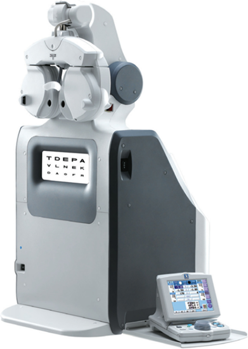
근거리시력 자동 포롭터(TS-310, NIDEK, Japen)를 이용하여 원거리 완전교정 상태에서 40cm용 근거리 시표를 사용하여 소수시력(DECI-MAL)으로 기록 하였다(Fig. 2). 근거리 대비감도 비교는 Good-Lite Company의 Adult Near Contrast Test(제품번호 587700)시표를 사용하였다. 완전교정 시험테를 착용하고 contrast 100%, 25%, 10% 시표로 측정하였으며, 3개의 대비감도 비교군에 따른 시력 변화의 차이를 비교 분석하였다.
자각적 조절력측정법은 Push-up, (−)렌즈 부가법(Minus lens to blur test)가 있으며,[23-24] OTUS-P는 렌즈를 교환하는 방식의 기기이기 때문에 (−)렌즈 부가법으로 측정하였다. 자동 포롭터(TS-310, NIDEK, Japen)를 이용하여 원거리 완전교정상태에서 측정하였다(Fig. 2).
안압측정은 리바운드 안압계 Icare Rebound Tonometer(Tiolat Oy, Helsinki, Finland) 자동안압검사기를 사용하여 측정하였다.[25-26]
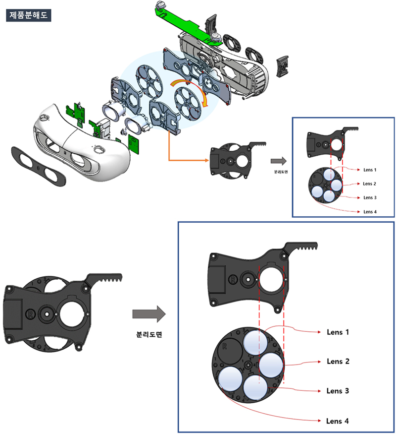
OTUS-P는 시기능 훈련(=Vision training=Vision theraphy) 기기로 분류되며, 플리퍼 타입의 훈련을 기반으로 조절용 이성과 조절력을 향상시킬 수 있는 기기이다. OTUS-P의 내부 구조는 Fig. 3에 제시되고 있는데, head mount 형태를 가지는 웨어러블 디바이스로써 내부에 구면렌즈를 장착한 리볼버 형태의 돌림판이 내장되며, 돌림판의 회전을 통해 눈에 적용되어 플리퍼 타입의 훈련이 이루어지도록하는 구조이다. 리볼버 형태의 돌림판 내부 렌즈들의 구성은 Table 3에 제시하였다.

Illustration showing the internal structure of the vision training device OTUS-P and the structure of the revolver-type lens wheel.
여기서 Lens1은 환자의 가입도(addition power) +0.50 D를 부과한 것이고, Lens2는 고정 굴절력 +2.00 D, Lens3은 환자의 가입도, Lens4는 가입도 −0.50 D를 부과한 것이다. 훈련 시 적용 렌즈 순서는 Lens1-Lens2-Lens3-Lens2-Lens4-Lens2이며, 이를 1 cycle로 하여 30 분간 반복 훈련한다.
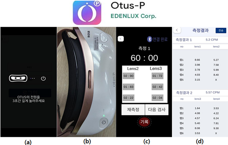
본 연구에서는 (주)에덴룩스에서 개발한 APP과 OTUS-P기기를 블루투스로 연결 후 Lens2(+2.00 D), Lesn3(최대 조절 자극렌즈)의 반응속도를 측정하여(Fig. 4 (c))반응속도가 기록되고 정보가 기기로 전송되도록 한다(Fig. 4(d)). 기기로 전송되는 데이터는 Lens2와 Lens3의 반응속도이며 측정된 반응속도대로 기기의 렌즈변경속도가 적용되어 훈련된다. Lens1과 Lens4의 렌즈 적용속도는 Lens3의 반응 속도와 동일하게 전송한다.

Stages upon activation of the OTUS-P application. (a) Presentation of the status of the Power input stage, (b) appearance of the device when the power button is pressed for 3 seconds, (c) display of the accommodative reaction speed measurement data by Lens2 and Lens3, and (d) appearance of the training data records.
(1) 분석 대상군 및 분석방법
본 임상시험의 대상자로부터 얻어진 자료의 분석군은 PPS(per protocol set)군으로 임상시험계획서대로 임상시험을 완료한 모든 피험자(1. 선정기준, 제외기준을 모두 충족시키는 환자. 2. 병용이 허용되지 않는 약물을 복용하지 않은 환자. 3. 최종방문시 1차 유효성 평가가 이루어진 환자)이다.
(2) 유효성 평가 변수에 대한 분석
Baseline 대비 4주 후 가입도 수치의 변화, 원거리 완전 교정 상태에서 근거리 시력 수치의 변화, 조절력((−)렌즈 부가법)수치의 변화, 대비감도 수치의 변화, 안압 전후의 변화를 기술통계량으로써 각 방문 시점별 평균, 표준편차, 중앙값, 최소값, 최대값 등을 제시하였다. 또한 분석방법으로써 해당 변수의 정규성을 검토한 후 정규성을 만족하면 Paired t-test를 시행하여 의료기기 적용에 따른 전후 차이를 비교 검정하였다.
(3) 안전성 평가 변수에 대한 분석-이상사례(adverse event, AE)
이상사례는 다음 항목으로, 1. 임상시험용의료기기에 대해 알려진 약리작용, 2. 임상적으로 유의성 있는 실험실검사의 이상치, 3. 이학적 검사에서 발견된 이상 반응, 4. 임상시험용의료기기나 같은 계열의 의료기기에서 이전에 관찰되었던 유사한 작용, 5. 유사한 의료기기와 관련 있다고 자주 보고된 반응, 6. 임상시험용의료기기 적용의 시간과 연관되어 나타나는 반응이다. 각각의 이상사례에 대해 한 번 이상의 이상사례를 경험한 피험자 수 및 백분율을 기술하였다. 이상사례의 정도, 시험군과의 관계, 조치 및 그 결과에 대해서 보고한 피험자 수 및 백분율을 기술하였다. 중대한 이상사례에 대해서는 시험군과의 인과 관계별로 발생 예 수(환자 수) 및 발생률을 제시하였다.
(4) 안전성 평가 변수에 대한 분석-활력징후
활력징후의 결과는 연속형 자료의 경우, 평균 표준편차, 중앙값, 최소값, 최대값을 정리하였고, 군내 의료기기 적용 전과 후의 차이는 정규성 검정을 만족하면 Paired t-test를 시행하였고 정규성 검정을 만족하지 않으면 Wilcoxon’s signed rank test를 시행하였다. 또한 의료기기 적용 전후의 정상범주를 벗어난 피험자들의 빈도수 및 백분율을 구하였다.
결과 및 고찰
1. 유효성 평가
PP(per-protocol) 분석군 결과를 살펴보면, 베이스라인 대비 4주 시점에서의 가입도 수치 변화량에 대해 우안(p=0.022)과 좌안(p=0.041)에서 모두 통계적으로 유의함을 나타내었다. 우안에서는 베이스라인에서 1.94(±0.38) D, 4주 시점에서 1.84(±0.51) D로서 변화량은 −0.1(±0.32) D로 확인되었고 좌안에서는 베이스라인에서 1.93(±0.35) D, 4주 시점에서 1.8(±0.46) D으로, 변화량은−0.1(±0.39) D로 확인되었다(Table 4).
PP(per-protocol) 분석군 결과를 살펴보면, 베이스라인 대비 4주 시점에서의 근거리 시력 수치 변화량에 대해 양안에서 통계적으로 유의함을 확인하였다(p<0.001). 양안에서 베이스라인에서는 0.64(±0.26), 4주 시점에서 0.74(±0.26)으로, 변화량은 0.1(±0.20)로 확인되었다(Table 5).
PP군 결과를 살펴보면, 베이스라인 대비 4주 시점에서의 자각적 조절력 수치의 변화량에 대해 양안에서 통계적으로 유의함을 확인하였다(p<0.001). 양안에서 베이스라인에서는 −1.94(±0.55) D, 4주 시점에서 −2.36(±0.76) D으로, 변화량은 −0.42(±0.69) D로 확인되었다(Table 6). 따라서 조절력은 0.42(±0.69) D 만큼 개선되었다고 볼 수 있다.
PP군 결과를 살펴보면, 베이스라인 대비 4주 시점에서의 대비감도 수치 변화량에 대해 contrast 100% 시표(p=0.3)에서는 통계적으로 유의함을 확인할 수 없었으나 contrast 25% 시표(p=0.003), 10% 시표(p<0.001)에서 모두 통계적으로 유의함을 확인할 수 있었다. 100% 시표에서는 베이스라인에서 0.81(±0.29), 4주 시점에서 0.86(±0.26)으로서 변화량은 0.05(±0.17)로 확인되었고 25% 시표에서는 베이스라인에서 0.68(±0.21), 4주 시점에서 0.74(±0.21)으로서 변화량은 0.07(±0.15)로 확인되었으며, 10% 시표에서는 베이스라인에서 0.6(±0.18), 4주 시점에서 0.66(±0.19)으로 나타나 변화량은 0.06(±0.13)로 확인되었다(Table 7).
PP군 결과, 베이스라인 대비 4주 시점에서의 안압의 변화량에 대해 우안(p=0.137)과 좌안(p=0.107)에서 모두 통계적으로 유의함을 확인할 수 없었다. 우안에서는 베이스라인에서 13.85(±2.45) mmHG, 4주 시점에서 14.43(±2.19) mmHG으로 나타나 변화량은 0.58(±2.81) mmHG로 확인되었고 좌안에서는 베이스라인에서 13.76(±2.57) mmHG, 4주 시점에서 14.34(±2.12) mmHG으로 나타나 변화량은 0.58(±2.56) mmHG로 확인되었다(Table 8).
2. 안전성 평가 결과
PPS 58명 중 임상의료기기 적용 이후에 한번 이상의 이상반응을 경험한 대상자는 1명으로 확인되었으며, 이는 시기능 훈련중 흔히 발생할 수 있는 두통이었다. 중증도는 경증이었으며 다른 치료없이 회복하였으며 후유증이 없는 것으로 확인되었다.
체온을 제외한 나머지 활력징후는 정규성을 만족하여 모수적인 방법으로 검정을 수행하였고 체온은 비모수적 방법인 Wilcoxon’s signed rank test를 실시하여 검정을 수행하였다. 혈압을 제외하면 나머지 시점에서는 통계적인 차이를 확인할 수 없었으며 맥박과 체온은 베이스라인 대비 모든 시점에서 통계적 차이를 확인할 수 없었다. 각 시점 별 기술통계량은 하기 표를 참고하였다(Table 9).
3. 고찰
시기능 훈련에는 Push-up, Flipper, Loose lens rock, Hart chart 등이 있는데, 이러한 방법들은 대부분 시기능 훈련센터 등을 방문해서 특정 시표 또는 그에 상응하는 크기의 활자가 있는 신문 또는 책 등을 주시하면서 훈련을 받도록 되어있다. 이 중에서 Push-up 또는 Flipper는 비교적 간단하여 홈트레이닝이 가능한 훈련으로 알려져 있지만, 렌즈의 교환이 수동적인 방법으로 설계되어 있다.[6,14-19] 본 연구에서 사용된 OTUS-P는 플리퍼 타입의 웨어러블 훈련기기로 홈트레이닝으로 진행되며, 렌즈의 자동교환으로 기존의 수동 렌즈 교환 방식의 수고를 덜어주며, 주시 물체를 핸드폰, 책 등으로 할 수 있도록 하여 사용자의 접근성과 편의성을 높혔다.
4주간 유효성 평가 변수의 변화량을 보았을 때 안압과 대비감도(100% 시표)를 제외하면 통계적으로 유의미함을 확인할 수 있었으며, 안전성에 대해서도 이상 없는 것으로 판단된다. 다만, 시기능 훈련의 특성상 4주의 훈련은 다소 짧은 기간으로, 지속적인 훈련을 진행한다면 유효성 평가 변수들의 개선량의 증가와 안압 및 대비감도(100% 시표) 또한 통계적인 유의미함을 가질 것으로 사료된다.[6,14-19]
결 론
본 연구는 가입도가 +0.75 D 이상인 노안환자(presbyopia) (45.62(±3.7)세, 남성 19명, 여성 41명)를 대상으로 플리퍼타입 훈련을 기반으로 한 자동화 및 홈트레이닝 시기능 훈련기구인 OTUS-P를 사용하여 하루 30분 적용하였으며, Baseline 대비 4주차의 유효성 평가 변수를 측정하여 차이를 분석하였다. 그 결과 4주 후 가입도는 우안 0.1(±0.32) D, 좌안 0.1(±0.39) D 만큼 감소되었고, 근거리 시력은 0.1(±0.20) 만큼 개선되었고, 자각적 조절력은 0.42(±0.69) D 만큼 개선되었고, 대비감도는 25% 시표에서 0.07(±0.15), 10% 시표에서 0.06(±0.13) 만큼 개선되었으며, 안압은 우안 0.58(±2.81) mmHG 만큼, 좌안 0.58(±2.56) mmHG 만큼 상승하였으나 정상범위 이내에서의 변화로 안전성에 문제가 없다고 판단된다. 그러나 안압의 경우 우안(p=0.137), 좌안(p=0.107)으로 나타나 통계적으로 유의미하지 않았으며, 이는 기간을 늘여 임상을 진행하면 통계적으로 유의미한 값을 가질 것으로 판단된다. 안전성 평가결과, 이상반응, 활력징후에서 임상적으로 유의미한 변화는 없어 안전하다고 판단된다.
4주간의 비교적 짧은 훈련 기간이지만 조절훈련을 통해 노안 개선에 도움이 되는지 확인할 수 있었다. 따라서, 향후 연구에서는 12주 이상 훈련을 적용하고 이에 따라 예상되는 문제점인 렌즈 범위의 제한과 훈련 방법의 다양성을 개선하여 상승효과를 확인하고자 한다.
Acknowledgments
본 연구는 정부(과학기술정보통신부,산업통상자원부,보건복지부,식품의약품안전처)의 재원으로 범부처전주기의료기기연구개발 사업단의 지원을 받아 수행된 연구임(과제고유번호 : RS-2020-KD000285)
References
- Plainis S, Charman WN, Pallikaris IG. The physiologic mechanism of accommodation. Cataract and Refractive Surgery Today Europe. 2014;4:23-28.
-
Kwon JW. What is presbyopia?. J Korean Med Assoc. 2019;62(12):608-610.
[https://doi.org/10.5124/jkma.2019.62.12.608]

-
Kwon KI, Kim HJ, Park M, et al. The functional change of accommodation and convergence in the mid-forties by using smartphone. J Korean Ophthalmic Opt Soc. 2016;21(2):127-135.
[https://doi.org/10.14479/jkoos.2016.21.2.127]

-
Almutairi MS, Altoaimi BH, Bradley A. Accommodation and pupil behaviour of binocularly viewing early presbyopes. Ophthalmic Physiol Opt. 2017;37(2):128-140.
[https://doi.org/10.1111/opo.12356]

-
Wajuihian SO. Frequency of asthenopia and its association with refractive errors. Afr Vis Eye Health. 2015;74(1):a293.
[https://doi.org/10.4102/aveh.v74i1.293]

-
Kim NS, Lee HM. Changes in accommodative function in patients in their 30s and 40s by visual function training. J Korean Ophthalmic Opt Soc. 2023;28(4):341-351.
[https://doi.org/10.14479/jkoos.2023.28.4.341]

-
Wolffsohn JS, Davies LN. Presbyopia: effectiveness of correction strategies, Prog Retin Eye Res. 2019;68:124-143.
[https://doi.org/10.1016/j.preteyeres.2018.09.004]

-
Choi KU, Lee MH, An Y. Investigation of aging awareness and eye health care of early presbyopia. Korean J Vis Sci. 2023;25(2):157-167.
[https://doi.org/10.17337/JMBI.2023.25.2.157]

- KOSIS(Statistics, Korea), Korean Statistical Information Service: Population situation board, 2024. https://kosis.kr/visual/populationKorea/PopulationDashBoardMain.do, (04 January 2024).
-
Park JH, Kim MJ. Surgical treatment of presbyopia. J Korean Med Assoc. 2014;57(6):520-524.
[https://doi.org/10.5124/jkma.2014.57.6.520]

-
Katz JA, Karpecki PM, Dorca A. et al. Presbyopia – a review of current treatment options and emerging therapies. Clin Ophthalmol. 2021;15:2167-2178.
[https://doi.org/10.2147/OPTH.S259011]

-
Kannarr S, El-Harazi SM, Moshirfar M, et al. Safety and efficacy of twice-daily pilocarpine HCl in presbyopia: the virgo phase 3, randomized, double-asked, controlled study. Am J Ophthalmol. 2023;253:189-200.
[https://doi.org/10.1016/j.ajo.2023.05.008]

-
Gualdi L, Gualdi F, Rusciano D, et al. Ciliary muscle electrostimulation to restore accommodation in patients with early presbyopia: preliminary results. J Refract Surg. 2017;33(9):578-583.
[https://doi.org/10.3928/1081597X-20170621-05]

-
Hwang HY, Cho HG. Changes of addition by accommodative training on early presbyopia. Journal of the Korea Academia-Industrial cooperation Society. 2010;11(6):2190-2195.
[https://doi.org/10.5762/KAIS.2010.11.6.2190]

-
Wick B. Vision training for presbyopic nonstrabismic patients. Optom Vis Sci. 1977;54(4):244-247.
[https://doi.org/10.1097/00006324-197704000-00009]

-
Liza SJ, Choe S, Kwon OS. Testing the efficacy of vision training for presbyopia: alternating-distance training does not facilitate vision improvement compared to fixed-distance training. Graefes Arch Clin Exp Ophthalmol. 2022;260(10):1551-1563.
[https://doi.org/10.1007/s00417-021-05548-8]

-
Sterkin A, Levy Y, Pokroy R, et al. Vision improvement in pilots with presbyopia following perceptual learning. Vision Res. 2018;152:61-73.
[https://doi.org/10.1016/j.visres.2017.09.003]

- Hwang HY. A study on accommodative training of initial presbyopia aged in 40s. MS Thesis. Kyungwoon University, Gumy. 2009;3-50
- Calef T. Portland presbyopia onset delay study. PhD Thesis. Pacific University, Oregon. 1995;4-21.
- Lee BR. A study on the legal problems and improvements of the occupational range of the occupants. Law Review. 2019;19(3):477-501.
-
Joo SH, Shim MS, Shim JB. The study on change of refractive error and addition in progressive eyeglasses lens wearers. J Korean Ophthalmic Opt Soc. 2013;18(4):399-404.
[https://doi.org/10.14479/jkoos.2013.18.4.399]

- Kim CJ, Kim HY, Kim JM. Comparison of contrast sensitivity at near between functional progressive addition lenses and sigle vision lenses. J Korean Ophthalmic Opt Soc. 2010;15(4):381-388.
-
Momeni-Moghaddam H, Kundart J, Askarizadeh F. Comparing measurement techniques of accommodative amplitudes. Indian J Ophthalmol. 2014;62(6):683-687.
[https://doi.org/10.4103/0301-4738.126990]

- Kim HM, Son JS, Kim IS, et al. Comparison of accommodative amplitude based on occupation of initial presbyopia. J Korean Ophthalmic Opt Soc. 2008;13(4):135-139.
-
Lee K, Lee JY, Moon JI, et al. Comparison of icare rebound tonometer with goldmann applanation tonometry. J Korean Ophthalmol Soc. 2013;54(2):296-302.
[https://doi.org/10.3341/jkos.2013.54.2.296]

-
Lee KS, Kim SK, Kim EK, et al. Comparison of intraocular pressure measured by non-contact tonometer, rebound tonometer, tono-pen, and goldmann applanation tonometer. J Korean Ophthalmol Soc. 2014;55(1):47-53.
[https://doi.org/10.3341/jkos.2014.55.1.47]


